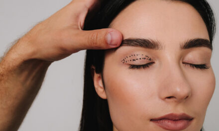
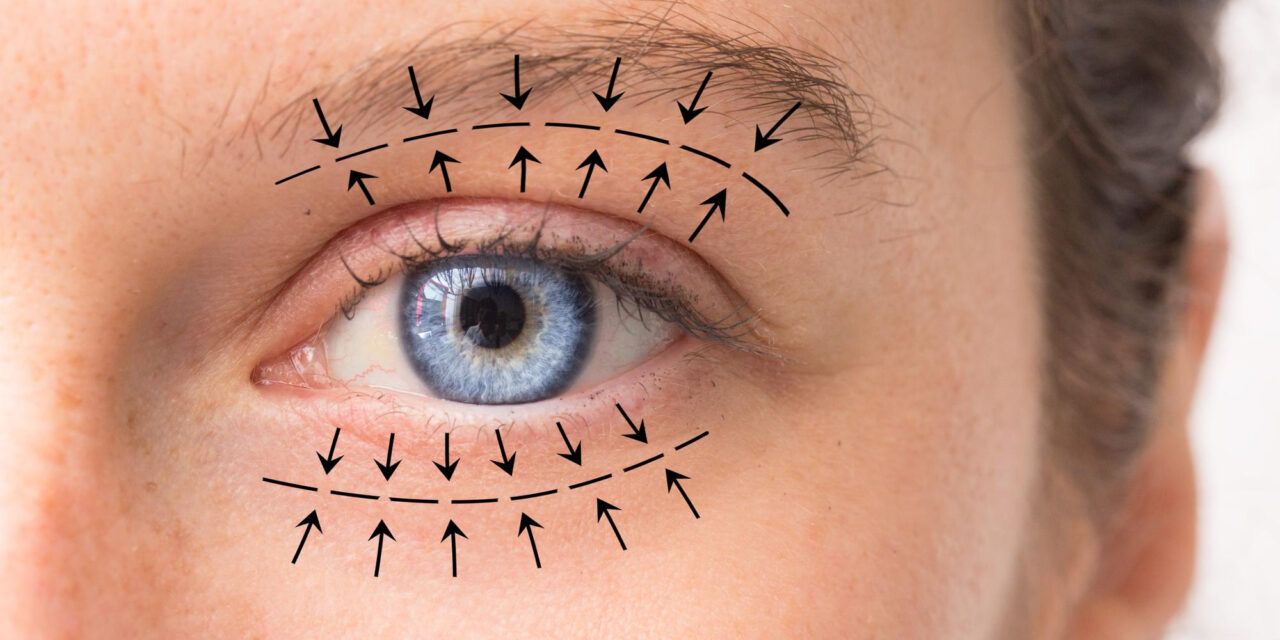
Anyone researching blepharoplasty risks is usually asking two questions at once: what will normally happen after eyelid surgery, and what could go wrong in a way that matters? This guide answers both. Blepharoplasty (eyelid surgery, sometimes called an eye lift) removes or repositions skin, muscle and fat around the eyelids. Because the eyelids protect the eye, even small changes in how they move, close or sit against the eyeball can affect comfort and, rarely, vision. That is why the risk conversation for eyelid surgery deserves more care than the casual “swelling and bruising” summary found on many clinic pages.
Below you will find the expected short-term effects, the complications that are uncommon, the very rare ones that are emergencies, the personal factors that raise or lower risk, and a practical pathway for reducing it. Everything is general education for U.S. readers, not medical advice, and it cannot tell you what your own risk is. Only an in-person evaluation can do that.
A note on vocabulary used throughout: “upper blepharoplasty” means surgery on the upper lids, “lower blepharoplasty” means surgery on the lower lids, and “four-lid” or “combined” surgery means both. “Complication” means an unplanned problem that goes beyond the normal healing course, while “expected effect” means something that most people experience and that usually fades. The first major section explains why that line matters and why it is not always sharp.
Several related guides on this site go deeper on single topics. The complete guide to eyelid surgery covers the procedure as a whole, the upper eyelid risks guide narrows in on upper-lid surgery, and the eyelid surgery recovery timeline walks through healing week by week. This page is the safety hub that ties them together.
Understanding blepharoplasty risks: terms, evidence and what “safe” can honestly mean
Risk talk goes wrong in two predictable ways. Some pages bury it under reassuring language, and others list every conceivable problem with no sense of proportion. Neither helps a person deciding whether to have surgery. This first section sets the ground rules for the rest of the guide: which words mean what, how much the published numbers can really tell you, and what it means, and does not mean, to say an operation is safe.
The vocabulary of risk
A few terms recur in consent forms and clinic handouts. Understanding them in advance makes the conversation with a surgeon far more productive, because you can tell when an answer is specific and when it is evasive.
Side effects, complications and adverse outcomes are three different things
A side effect or expected effect is something most patients experience after the operation and that usually settles without treatment. After eyelid surgery that list is familiar: swelling, bruising, a gritty or dry feeling, tearing, tightness, light sensitivity and some blurring while ointment coats the eye. The American Society of Plastic Surgeons (ASPS) describes swelling, bruising, irritation, dry eyes and discomfort as part of ordinary recovery, to be managed with medication, cold compresses and ointment.
A complication is a problem that is not part of the planned course of healing. It may be minor and temporary, such as a small stitch reaction, or major and lasting, such as an eyelid that no longer closes fully. The ASPS safety page for eyelid surgery lists, among others, anesthesia risks, bleeding, infection, numbness, difficulty closing the eyes, dryness, outward-rolling of the lower lid (ectropion), lid lag, persistent pain, light sensitivity, unfavorable scarring, the possible need for revision, and temporary or permanent vision changes with a very rare risk of blindness. Mayo Clinic offers a similar list and adds eye muscle injury and skin discoloration.
An adverse outcome is a broader phrase. It can include a technically uncomplicated operation that still leaves the person unhappy, for instance because the result looks different from what they expected, or because the problem they hoped to fix (under-eye darkness, for example) was not something eyelid surgery addresses. These disappointments are not always “complications” in the medical sense, but they are real, and a good consent conversation covers them.
The line between an expected effect and a complication is not always crisp. Mild lid-height asymmetry in the first weeks can be ordinary swelling. The same asymmetry at six months may be a complication. Dryness for several days is expected; dryness that damages the corneal surface is not. Timing, severity and trajectory are what separate the categories, and the next section returns to that idea in detail.
Where the numbers come from, and why they differ
People often want one percentage, and the honest answer is that no single figure applies. Published numbers vary because studies differ in what they count, who they include and how long they follow patients. Three examples show why.
First, a 2004 survey of members of the American Society of Ophthalmic Plastic and Reconstructive Surgery asked surgeons to report vision-threatening bleeding after cosmetic eyelid surgery. The abstract reports 237 responding surgeons representing 269,433 cosmetic eyelid cases, with orbital hemorrhage at roughly 0.055 percent (about 1 in 2,000) and permanent visual loss at roughly 0.0045 percent (about 1 in 22,000). That is the largest dataset this guide could verify, but it relies on surgeons’ recollection and reporting, it is more than twenty years old, and it addresses one specific complication only.
Second, a single-center retrospective chart review in the Brazilian Journal of Otorhinolaryngology followed 200 consecutive patients who had transcutaneous blepharoplasty between 2007 and 2009. It recorded some kind of complication in 9.5 percent of patients (19 of 200), with lower-lid malposition in 3 percent and chemosis (swelling of the clear membrane over the white of the eye) in 6 percent. The authors counted minor problems, which explains the higher overall figure, and the results reflect one team, one technique and one patient group.
Third, review articles such as those by Lelli and Lisman (Plastic and Reconstructive Surgery, 2010) and by the authors of a 2006 review in the journal Orbit describe the spectrum of complications and how to prevent them but do not claim to give a universal rate. The Orbit review characterizes post-blepharoplasty complications as usually infrequent, minor and transient, while noting that the most serious can threaten sight.
It also helps to know what kind of evidence sits behind each number. Surveys of surgeons capture rare events across large volumes but depend on recall. Single-center chart reviews are detailed but reflect one team’s habits and patients. Narrative reviews synthesize experience and teach prevention, but they are shaped by what their authors have seen. Prospective studies, like the two small 2021 tear-film studies discussed later in this guide, measure specific outcomes carefully but usually in narrow groups over weeks or months. None of these is a registry that follows every eyelid operation in a region, and this review did not find one. In evidence-hierarchy terms, most of what is known about blepharoplasty complications rests on case series, expert reviews and small prospective studies, and the honest consequence is that confident claims about exact rates are rarely justified. What the literature does support, consistently, is the catalog of complications, their mechanisms, the factors that make them more likely, and the value of recognizing them early. Those are the points on which the sources agree, and they are the ones this guide leans on.
The takeaway is practical. When a website quotes a precise complication percentage without saying where it came from, treat it with suspicion. When a surgeon quotes a figure at consultation, a fair follow-up is to ask whether it comes from published literature or from their own cases, and over what period. This guide avoids numbers it cannot trace to a source, and it flags the few it uses with their year and population.
Is blepharoplasty safe? Putting blepharoplasty risks in context
“Is blepharoplasty safe?” is one of the most common questions people type, and it deserves a straight answer that avoids both alarm and reassurance.
What “safe” can and cannot mean for an elective operation
Elective surgery is never free of risk. Anesthesia carries risk, any incision can bleed or become infected, and the eyelids sit directly in front of an organ that does not tolerate surprises. When a clinic says a procedure is “safe,” the claim is relative and conditional: safe enough for an appropriately selected patient, performed by a trained surgeon, in a suitable facility, with proper anesthesia and follow-up. Change one of those conditions and the picture changes.
The reasonable summary supported by the sources reviewed here is that most people who have eyelid surgery recover without a serious problem, that minor and temporary issues are common, that a smaller group experience complications that need treatment or revision, and that a very small number face sight-threatening events. Those categories are described in the medical literature; precise frequencies depend on the study. A procedure being common does not make it trivial, and a risk being rare does not make it irrelevant to the person it happens to.
Another distinction matters: the risks of a procedure are not the same as the risks to you. A person with thyroid eye disease, significant dry eye disease, or a bleeding disorder is not walking into the same risk profile as a person with none of those, even if both are having “the same” upper-lid operation. Later sections take up those personal factors one by one.
Consider two hypothetical people, described only to show how the factors interact; they are not real patients. The first has hooded upper lids from skin laxity, healthy tear production and normal blood pressure, takes no blood thinners, and is planning upper-lid surgery alone with local anesthetic and light sedation given by a separate anesthesia professional. The second has lax lower lids, a history of dry eye and blood pressure that is sometimes elevated, takes a daily aspirin, and is considering four-lid surgery under general anesthesia in a setting he has not yet asked about. Both could be told that blepharoplasty is safe, and both could have good outcomes. But the questions each needs answered before saying yes are not the same, and the second person has more conversations to have, with more clinicians, before the decision is sound. That does not mean the second person should be refused surgery. It means risk belongs to the plan and the person together, not to the operation’s name.
Four levers that move risk: the procedure, the person, the team and the setting
It helps to organize everything that follows around four levers.
The procedure lever covers which lids are operated on, what is removed or repositioned, how the incision is made (through the skin or inside the lid), and whether anything else is done at the same time, such as a brow lift, ptosis repair, laser resurfacing or fat grafting. More tissue manipulation and more simultaneous work generally mean more variables.
The person lever is anatomy and health: eyelid laxity, how prominent the eyes are, tear production, thyroid status, blood pressure, bleeding tendency, medications and supplements, smoking, scar tendency, and expectations.
The team lever is the training and judgment of the surgeon and the anesthesia professional, and how the group handles problems. Many complications are linked to planning errors, such as removing too much skin or missing pre-existing lid laxity, rather than to bad luck, which is why the literature stresses careful pre-operative assessment.
The setting lever covers the facility and its emergency readiness, the type of anesthesia, monitoring, and the quality of post-operative instructions and access to the surgical team afterward.
The map below summarizes how common effects, less common complications, serious complications and personal risk factors fit together, and why a discussion of risk should end with “what changes the plan” rather than a generic list.
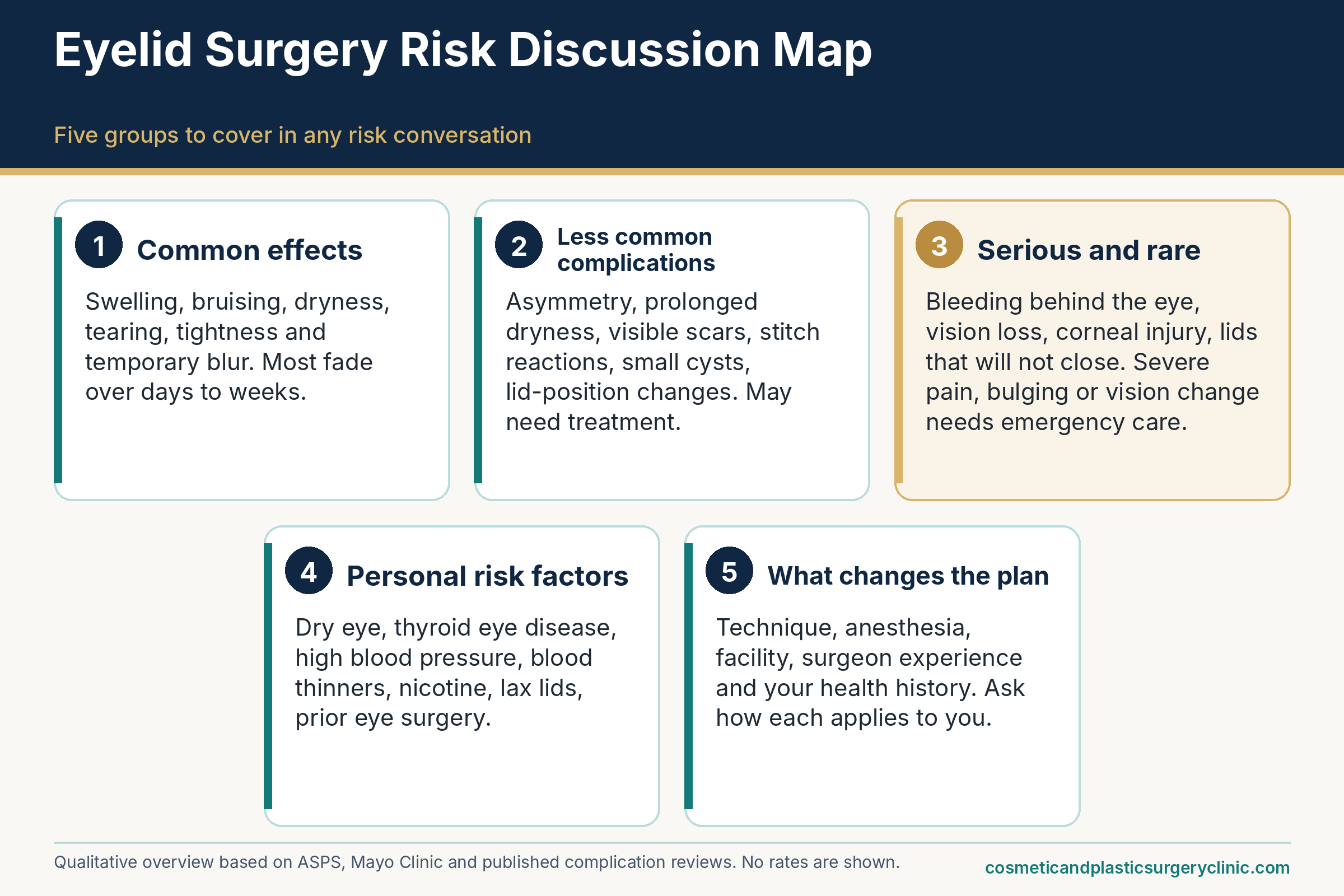
| Tier | Examples | Usual timing | Typical next step | Evidence note |
|---|---|---|---|---|
| Expected effects | Swelling, bruising, tightness, dryness, tearing, light sensitivity, ointment-related blur | Peak in the first days; fade over weeks | Follow written aftercare; routine follow-up | Described by ASPS, Mayo Clinic and MedlinePlus; duration varies by person |
| Less common complications | Asymmetry, prolonged dryness, small cysts (milia), stitch reactions, noticeable scars, chemosis | Days to months | Call the surgical team; conservative treatment first in many cases | Reviews describe these without a universal rate; single-center series report them in a minority of patients |
| Complications needing procedural care | Lid retraction or ectropion, incomplete closure with corneal exposure, ptosis, infection, wound opening, double vision that persists | First weeks to many months | Prompt evaluation; sometimes a second procedure after healing | Frequency depends on technique, anatomy and surgeon; revision is sometimes needed |
| Rare emergencies | Bleeding behind the eye, sudden vision loss, severe one-sided pain with bulging | Often early, in the first hours to first day | Emergency evaluation without delay | 2004 ASOPRS member survey (abstract): about 1 in 2,000 for hemorrhage, about 1 in 22,000 for permanent visual loss |
Expected short-term effects after eyelid surgery, and where they cross a line
Most of what people worry about in the first week after eyelid surgery is normal. The skin around the eyes is thin and richly supplied with blood vessels and lymphatics, so it shows swelling and discoloration more dramatically than almost anywhere else on the body. Knowing what ordinary looks like is half the battle, because it lets you relax about the expected and notice the unexpected. This section separates the two, then describes the situations where an expected effect starts to look like a problem.
The first days: what usually happens
The first several days are when most of the visible and physical effects appear. Instructions differ from one surgeon to another, so treat what follows as a general description of the pattern, not as instructions for your own recovery.
Swelling and bruising
Swelling (edema) is the body’s response to surgical tissue handling, and it typically builds over the first day or two before easing. Eyelid swelling can make the eyes feel heavy, change how the lids sit, and temporarily alter the shape of the eye opening. Bruising (ecchymosis) can spread well beyond the incision lines, sometimes down the cheek or toward the temple, because blood tracks along tissue planes. Both effects are often strongest on the morning after surgery, particularly after lying flat, which is why many surgical teams recommend keeping the head elevated and using cold compresses in the early period.
How long it lasts varies. Mayo Clinic describes bruising and swelling diminishing over roughly ten to fourteen days, while the MedlinePlus encyclopedia entry on eyelid lift surgery says bruising can last two to four weeks. Those two sources are not in conflict. They describe different points on a spectrum, and individual healing, skin tone, medications, the extent of surgery and whether the lower lids were treated all shift where a particular person lands. Residual swelling, especially in the morning, can persist for weeks to months after the visible bruising is gone, and lower-lid swelling often takes longest to resolve.
Two patterns are more worrisome than diffuse swelling and bruising: swelling that is markedly greater on one side and increasing rather than leveling off, and swelling that comes with severe pain or a change in vision. Those are discussed in the red-flag section below. Lopsided swelling by itself can still be normal in the first couple of days, since people rarely sleep or bleed identically on both sides, but it is exactly the sort of thing to mention to the surgical team the same day, because only an examination can distinguish ordinary asymmetry from a collection of blood.
Dryness, tearing, light sensitivity and blurred vision
Eyelid surgery interrupts the usual mechanics of blinking and tear spreading for a while. The ASPS recovery page lists dry eyes among the normal early effects and recommends diligent sun protection and darkly tinted sunglasses until healing is complete. Mayo Clinic lists blurred vision, watering eyes and light sensitivity among the common temporary effects. Several things contribute at once.
Swollen lids may not close fully during blinking or sleep, which dries the surface. At the same time the eye can overproduce tears as a reflex response to irritation, so a person can have simultaneously dry and watery eyes. Lubricating ointment, which many surgeons prescribe in the first days, coats the surface and often blurs vision for a few minutes after application. That kind of blur clears with blinking and is different from blur that persists, worsens, or is accompanied by pain.
A small prospective study published in Frontiers in Medicine in 2021 offers some measured context. It followed 55 young women (110 eyes, ages 19 to 31) after cosmetic transcutaneous upper blepharoplasty and found changes in blinking one week after surgery, with reduced blink frequency and more incomplete blinks, that returned to baseline within a month. The authors found no clinical correlation between those blink changes and the ocular surface measures they tracked. The study is useful as an illustration of a temporary pattern, but it involved a narrow, young group having upper-lid surgery only. It does not describe what happens to older adults, people with existing dry eye, or people who have lower-lid surgery.
Sensation and appearance during healing
Beyond the first days, the more subtle changes can feel stranger than swelling because they involve sensation and the look of the lid margin. Most are expected, and most improve.
Tightness, numbness, itching and other odd sensations
A tight or pulling feeling in the lids is common, partly from swelling and partly from the new tension of the incision lines. Numbness along the lid or lashes also occurs because small sensory nerves are cut during surgery; the ASPS safety page lists changes in skin sensation, including numbness affecting the eyelashes, among possible risks. Sensation often returns gradually, though the time course varies and some patients notice small areas of altered feeling for months.
Itching is common as incisions heal and the skin recovers. It can be tempting to rub the eyes, but rubbing fresh incisions or a dry eye can damage tissue, so surgeons routinely advise against it and may suggest lubricating drops or ointment instead. Mild irritation at the incision sites is also described on the ASPS recovery page. Some people notice small, firm bumps along the incision as stitches dissolve or are removed; these are discussed with the other healing-related problems in a later section.
The sensations that deserve a call are not the mild ones but the severe, escalating or one-sided ones: pain that is out of proportion to what your team described, a deep ache behind the eye, or pressure that builds instead of easing. Those can point to bleeding or inflammation inside the orbit and are covered under red flags.
The temporary asymmetry stage, and how to tell expected from not expected
During healing, the two eyes rarely look alike. One side may swell more, one crease may look higher, one lower lid may sit slightly differently against the eyeball. Surgical reviews describe asymmetry in lid height and crease position as the most common thing patients notice, and a 2012 review in Plastic Surgery International recommends waiting at least three months before deciding on a touch-up for minor asymmetries or persistent drooping, so that swelling and scar maturation can settle. The review described the temptation to operate early as chasing the result.
That advice does not mean waiting through a true problem. A useful rule of thumb, offered here as general education only, is to compare three things against what your surgeon told you to expect: direction (is it improving, flat or worsening?), function (can you close the eyes comfortably, is vision stable?), and side effects that feel disproportionate (severe pain, fever, discharge). A temporary difference that is slowly narrowing, with comfortable closure and stable vision, fits the expected course. A difference that is widening, or one that comes with exposure symptoms or pain, belongs in a same-day call.
To make the pattern concrete, here is an illustrative composite of an ordinary early course, assembled from the general descriptions in the sources and not a prediction for any individual. In the first day or two, swelling builds, the lids feel heavy, and ointment makes vision hazy for a few minutes at a time; sleeping with the head elevated and using cold compresses are typical. By days three to five, bruising is usually at its most colorful and may have drifted below the eye. Sutures that need removal are commonly taken out within about a week, according to ASPS, and the incision lines may feel tight and itchy. Over the second week, bruising fades through green and yellow, and Mayo Clinic puts the usual window for swelling and bruising to diminish at roughly ten to fourteen days, while MedlinePlus allows two to four weeks for bruising. After that, what remains is mostly lingering morning puffiness, a slightly pink scar, and occasional dryness. A person whose course looks very different from this, in either direction, is not necessarily having a problem, but a difference worth noticing is a reason to ask.
Table 2 sets out common early experiences next to the point at which many surgical teams would want to hear from you. Your own team’s written instructions take priority over any general table.
| Experience | Often expected | Mention to the team promptly if | Needs urgent or emergency evaluation if |
|---|---|---|---|
| Swelling and bruising | Peaks in the first days; fades over about one to several weeks; can spread to the cheek | Much greater on one side, or still increasing after the first couple of days | Rapid one-sided swelling with severe pain, bulging or vision change |
| Dry, gritty or watery eyes | Common early; managed with the lubricants your team prescribes | Burning, redness or light sensitivity that is increasing, or the eye cannot close comfortably | Sharp eye pain with reduced vision |
| Blurred vision | Brief blur after ointment that clears with blinking | Blur that persists between ointment applications | Sudden or progressive loss of vision, or a dark area in the visual field |
| Tightness and numbness | Pulling sensation; patchy numbness along lid or lashes | Numbness spreading or accompanied by weakness elsewhere in the face | Any new neurologic symptoms such as facial drooping or confusion |
| Asymmetry | Common early; often narrows over weeks to months | Widening difference, or a lid that appears to sit too low or too high | Asymmetry plus sudden pain, bulging of one eye or inability to move the eye |
| Double vision | Brief doubling can occur early; Mayo Clinic lists it among temporary effects | Doubling that lasts or returns | Double vision with severe pain, bulging or vision loss |
One reason this distinction is hard for patients is that early recovery involves some anxiety, and anxious attention magnifies small differences. Taking dated, same-lighting photos at the interval your surgeon suggests can make trends visible and reduce the temptation to scrutinize the mirror hourly. It also gives the surgical team something concrete to review if you call. The photo habit is a practical aid, not a diagnostic test, and it should never delay a call about the red-flag symptoms described later.
Upper, lower and combined eyelid surgery: how the risk profile differs
“Eyelid surgery” covers several different operations that share a name. Removing a fold of excess skin from the upper lid is not the same undertaking as reshaping the lower-lid fat pads or tightening a lax lower eyelid. Each operation puts different structures at risk, and the complications that cluster around one are often rare in the other. Anyone comparing risk across clinics, or across websites, should start by asking which operation is being discussed.
Upper eyelid surgery
Upper blepharoplasty is the more common starting point and the one most often considered when excess skin weighs on the lid or blocks part of the visual field. The ASPS procedure page describes an incision placed within the natural crease of the upper lid, through which a surgeon can remove or reposition fat, tighten muscle and remove excess skin. A dedicated guide on this site covers upper eyelid surgery risks and complications in more depth; the sections here give the comparative picture.
What the upper-lid risk profile concentrates on
Three structures carry most of the upper-lid risk: the skin and muscle budget, the lid-lifting muscle (the levator), and the fat pads.
The skin budget matters because the upper lid has to close completely over the eye with every blink and during sleep. Taking too much skin can leave the lid unable to close fully, a problem called lagophthalmos, which dries the corneal surface. The 2012 Plastic Surgery International review describes leaving a defined amount of skin between the brow and the crease incision to help avoid this, and the StatPearls entry on lagophthalmos lists excessive tissue removal during blepharoplasty among the post-surgical causes of incomplete closure. Because skin removal is measured in millimeters, precision at the planning stage is critical.
The levator is the muscle and tendon-like sheet that lifts the upper lid. Injury, cautery or scarring near it can lower the lid’s resting height, producing ptosis (a drooping lid). Swelling or small bleeds can cause a temporary version that settles; the 2012 review indicates most postoperative ptosis resolves within about three months, while persistent cases may need repair. Surgeons guard against this by identifying the levator during dissection and by evaluating for pre-existing ptosis before surgery, since a lid that already sits low is a different problem from excess skin and calls for a different operation. The ASPS candidates page makes this distinction explicitly: ptosis, in which the lifting mechanism is stretched or weakened, requires a different surgical approach from standard cosmetic blepharoplasty.
The fat pads sit behind the orbital septum, and removing too much can leave the upper lid looking hollow or “sunken,” a point raised in the Brazilian series as a cause of premature aging of the orbit when fat is over-resected. Conservative fat management is a recurring theme in the literature for that reason. Other upper-lid issues include asymmetric crease height, a crease that is too high or too faint, and scars that are visible when the lid is raised or closed. The Orbit review of complications also lists pigmentation changes and over- or under-correction in its overview.
When upper-lid surgery is paired with ptosis repair or brow considerations
Lids and brows influence one another. When the eyebrow has descended, it can push extra skin onto the upper lid and mimic the heaviness of true lid excess. The ASPS candidates page notes that apparent eyelid drooping may come from forehead skin laxity or brow descent rather than from the eyelids alone. In that situation, removing lid skin without addressing the brow can disappoint, and in some cases the brow may sit lower afterward. The Brazilian review discusses brow ptosis as a complication area that is best handled by pre-operative assessment, patient education and, where appropriate, discussion of brow repositioning options. A separate guide covers the brow lift as its own operation.
Ptosis repair is also sometimes combined with blepharoplasty. The MedlinePlus article on eyelid lift surgery describes surgery that combines tightening of the lid’s lifting mechanism with skin removal when drooping obstructs vision. Combining the two increases the number of structures the surgeon is adjusting at once, and the more precise the lid-height planning must be. Patients with a history of dry eye, a high-set brow, or lids that already show retraction deserve a particularly careful discussion about how much change is wise.
Lower eyelid and combined surgery
The lower lid is structurally more delicate than the upper. It must hold a stable position against the globe, and it relies on a balance between its skin, its supporting muscle and its horizontal tightness. Disrupting that balance is the central worry in lower-lid surgery, and it explains why lower-lid complications attract so much attention in the literature.
Transcutaneous versus transconjunctival approaches: what differs and what the evidence cannot settle
The ASPS procedure page describes two basic ways to reach the lower-lid tissues. In the transcutaneous approach (often called subciliary), an incision runs just below the lash line, which allows excess skin to be removed and fat to be managed. In the transconjunctival approach, the incision is made on the inside surface of the lower lid, which allows fat to be removed or redistributed without cutting through the skin and does not by itself remove skin.
The difference has risk implications. Disturbing the outer layer of the lid, either by cutting through the skin and muscle or by removing too much skin, is a recognized route to lower-lid retraction (the lid margin pulling down to expose white of the eye, called scleral show) and ectropion (outward rolling). The Brazilian series attributed lower-lid malposition to excessive skin resection, scarring of the orbital septum and failure to anchor the lateral canthus, and it reported malposition in 3 percent of its transcutaneous patients. A Healio clinical discussion of managing lower-lid malposition lists horizontal laxity, excessive skin removal, globe prominence and scarring as the root causes. A transconjunctival approach avoids a skin incision and so, in principle, avoids some of those mechanisms, which is why many surgeons favor it when the main issue is fat bulging without much extra skin.
The evidence for declaring one approach better is more limited than marketing suggests. This review did not locate a verified head-to-head comparison that could responsibly be summarized, and the older conference reports that circulate online contain figures that are hard to interpret without full methods. What can be said is that the choice depends on anatomy: patients with excess skin, laxity, or a need for skin tightening may need an approach that addresses skin, while those with fat prominence and good skin quality may be candidates for an internal approach, sometimes paired with a peel or laser for skin quality, as the ASPS procedure page mentions. The risk of each approach is also modified by whether the lid is tightened horizontally at the same time. A fuller technical comparison lives in the guide to lower eyelid surgery techniques, and the lower eyelid candidacy guide explains who is evaluated for which approach.
Four-lid surgery, fat repositioning, canthal support and add-ons
Doing all four lids in one session is common. The advantage is a single recovery; the trade-offs are a longer operation, more cumulative swelling, and compounding of risks that affect eye closure. A shortened upper lid and a retracted lower lid together can make complete closure much harder than either would alone. This is one reason surgeons plan the whole eyelid complex rather than each lid separately, and why many ask patients to be conservative with skin removal. The guide to combining eyelid surgery with other procedures explores the trade-offs of adding a brow lift, facelift or resurfacing.
Several techniques are added to lower-lid surgery specifically to reduce the malposition risk or improve contour. Canthopexy or canthoplasty tightens or anchors the outer corner of the lid to the bone, and the Brazilian series describes it as a routine component of its lower-lid approach, with special attention for older patients with laxity, deep-set sockets or a negative vector (where the cheekbone sits behind the front of the eyeball). Fat repositioning moves rather than removes orbital fat to smooth the junction between lid and cheek, and its main trade-off is added technical complexity. Laser or chemical peel may be offered for skin quality; the 2006 Orbit review notes that newer laser procedures carry their own risks, including ectropion and burns. Filler in the tear trough is a separate nonsurgical choice with its own side effects, covered in the guide to under-eye filler side effects.
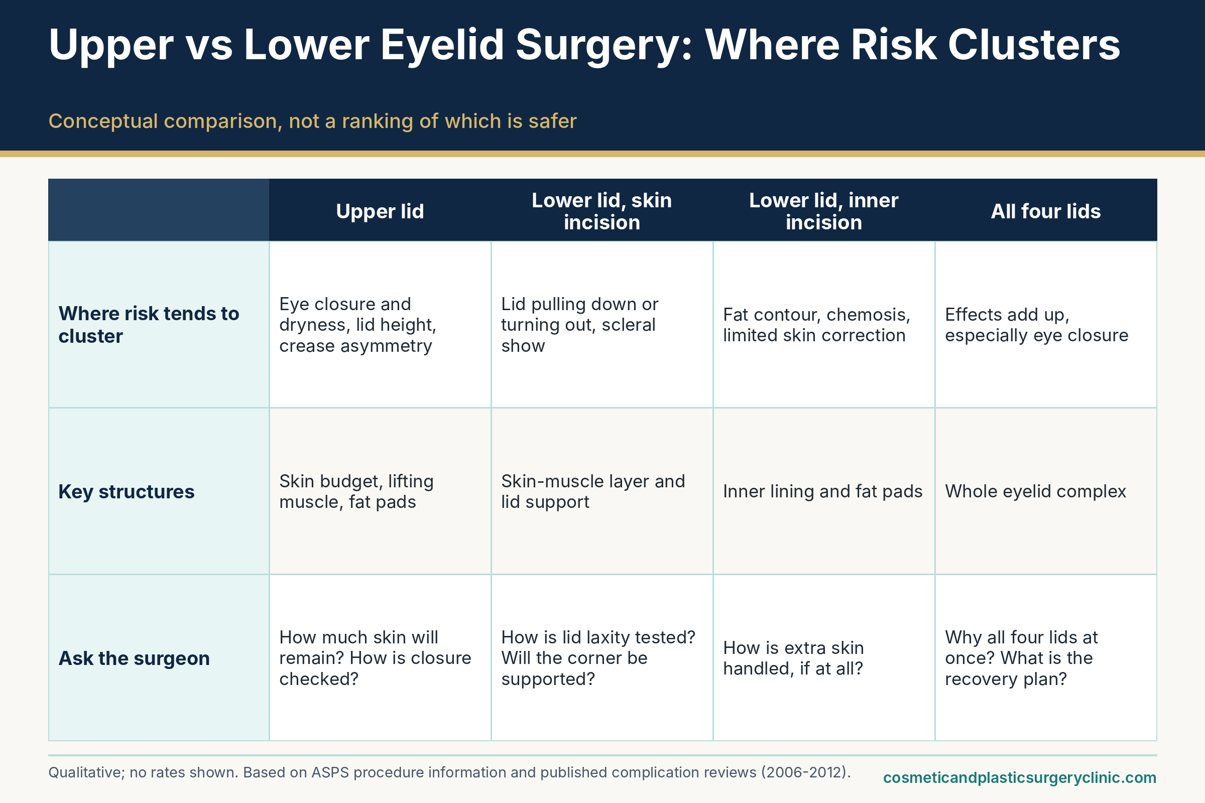
Table 3 summarizes where the risks tend to concentrate in each operation. It is a conceptual comparison drawn from the review literature, not a ranking of which is safer, because the operations are chosen for different anatomy and goals.
| Operation | Structures most at issue | Complications that tend to be discussed | Why they cluster there |
|---|---|---|---|
| Upper blepharoplasty (skin, with or without fat or muscle) | Skin budget, levator, fat pads, crease position | Incomplete closure and dry eye, ptosis, crease asymmetry, hollowing, visible scar | Upper lid must close fully; levator lies close to the plane of dissection |
| Lower blepharoplasty, transcutaneous (skin incision) | Skin-muscle layer, lid margin support, septum | Lid retraction, scleral show, ectropion, rounded outer corner, visible scar | Disturbing or shortening the outer layer pulls the lid down; lax lids compound the effect |
| Lower blepharoplasty, transconjunctival (internal incision) | Conjunctiva, fat pads, septum | Chemosis, fat-contour irregularity, limited skin correction, possible need for a skin adjunct | No external skin incision, but skin excess is not addressed by this route alone |
| Four-lid or combined with brow lift, ptosis repair or resurfacing | Whole eyelid complex plus adjacent structures | Cumulative swelling, combined effects on closure, longer anesthesia, more complex revision if needed | More structures adjusted in one session; effects can add up |
The figure below lays out the same comparison visually, as a quick reference to bring to a consultation.
A final point on this comparison: the operation’s name on a quote does not tell you how it will be done. “Lower blepharoplasty” might mean a transconjunctival fat adjustment, a skin-pinch excision, a skin-muscle flap with a canthal tightening, or a combination. Asking for the specific technique and the reason it was chosen for your anatomy is a core consultation question, and it appears again in the question tables later in this guide. Readers who want the broader procedural picture can use the overview of eyelid surgery techniques alongside this page.
Dry eye, exposure and lid-position problems
If you asked oculoplastic surgeons which eyelid surgery complications they think about most, the answer would probably not be the dramatic ones. It would be the quieter problems that arise when an eyelid no longer protects and lubricates the eye as well as it did before: a surface that dries out, a lid that will not close completely, or a lower lid that sits a little too low. These are among the most important blepharoplasty complications to understand, because they are more likely than the rare emergencies, they can be slow to resolve, and they are often preventable with careful planning.
The ocular surface: dryness, closure and corneal protection
The front of the eye depends on a thin, stable tear film, renewed with every blink, and on lids that close completely at night. Anything that changes blinking, closure or tear distribution can disturb that system. Surgery on the lids does exactly that, at least temporarily.
Dry eye after surgery, and why existing dry eye changes the calculation
Dry eye disease occurs when the eye does not make enough tears or when tears evaporate too quickly. The National Eye Institute describes symptoms as scratchiness, stinging, redness, light sensitivity and blurred vision, lists higher risk in people over 50, women, contact lens wearers and people with autoimmune conditions, and outlines management that ranges from over-the-counter artificial tears and lifestyle changes to prescription drops and tear-duct plugs. Mayo Clinic’s patient guide to blepharoplasty tells patients to discuss dry eyes with the surgeon before surgery, and ASPS lists dry eyes among the recognized risks.
Why would surgery worsen dryness? Several mechanisms are plausible contributors. Swelling and tightness can reduce the completeness of blinks. Removing skin or muscle can change how the lids meet the eye. Cutting small nerves affects sensation, which can alter the reflex that triggers tearing and blinking. And any disturbance of the oil-producing glands along the lid margin can destabilize the tear film. In most people with healthy eyes the effects are temporary: the 2021 prospective study of 55 young women, noted earlier, found blink changes that returned to baseline within a month after upper blepharoplasty.
The picture is less reassuring for people who already have dry eye disease. A separate 2021 Frontiers in Medicine study followed 20 people (40 eyes) after upper blepharoplasty, comparing those with and without pre-existing dry eye, with checks at one, three and six months. In people without prior dry eye, tear stability measures were shortened at one month and returned to baseline by six months. In the dry eye group, tear film instability and inflammatory markers in tears persisted through six months. The authors concluded that pre-existing dry eye is a risk factor for worse and more persistent ocular surface damage after upper blepharoplasty. The sample was small, and a laboratory-style study of tear markers does not tell you how a particular patient will feel, but the direction of the finding is consistent with the guidance surgeons give.
That consistency shows up in the guidance on LASIK, a procedure that itself reduces corneal sensation and can cause dry eye. A summary published on OphthalmologyWeb of a 2007 Plastic and Reconstructive Surgery paper by Korn and colleagues advised delaying eyelid surgery for at least six months after LASIK, evaluating dryness with formal testing, treating persistent dry eye before operating, and keeping fat removal conservative and avoiding removal of the circular eyelid muscle when surgery proceeds. That advice reflects expert opinion drawn from a single article, so it should not be read as a universal rule, but it illustrates how seriously ocular surface health is treated in pre-operative planning. Anyone who has had LASIK or other refractive surgery should say so at the first visit, and anyone who wears contact lenses should ask how long they will need to stay out of them afterward; one MedlinePlus description of typical instructions mentions avoiding contact lenses for about two weeks, but instructions vary.
Lagophthalmos, exposure keratopathy and corneal abrasion
Lagophthalmos means incomplete closure of the eyelids. The StatPearls entry on the condition explains why it matters: a full lid closure with a normal blink reflex is necessary to maintain a stable tear film and a healthy ocular surface. When closure is incomplete, the cornea is exposed, tears evaporate, and complications can progress from dryness to abrasions, persistent epithelial defects, ulcers and infection of the cornea (microbial keratitis), which can scar and affect vision. Excessive tissue removal during blepharoplasty is listed among the post-surgical causes.
Mild, temporary lagophthalmos is common in the early days because swelling stiffens the lids. Surgeons often ask patients to use ointment and, if needed, to tape or protect the eyes at night, and they check closure at follow-up. What matters is the trajectory. If closure gets easier as swelling subsides, it is likely tied to edema. If it stays incomplete after the swelling is gone, the cause may be a true shortage of skin, scarring, or a lid-position issue that needs a more specific plan. The 2012 review in Plastic Surgery International calls corneal exposure from inadequate closure one of the most serious blepharoplasty complications and describes progression to chronic exposure keratitis in severe cases, with management ranging from lubrication and bandage contact lenses to surgical lid elevation or grafting.
Corneal abrasion, a scratch on the clear front surface of the eye, can happen during surgery or in the early recovery period. The review by Lelli and Lisman places corneal abrasions in the early postoperative window, within the first week, and the 2012 review recommends protecting the eye with lubrication during placement and removal of protective shields and checking visual acuity and the front of the eye on the first postoperative visit. For patients, the practical meaning is that a sudden sharp pain, a sense of something stuck in the eye, light sensitivity that suddenly worsens, or tearing with a foreign-body feeling should be reported the same day rather than endured. Most simple abrasions heal quickly when treated, as the review notes for non-infected abrasions, but a missed one can turn into something more serious.
Prevention begins before the operation. Surgeons evaluate lid closure, tear production and blink quality in the consultation, avoid removing more skin than the lid can spare, and consider whether the combination of upper and lower lid surgery will leave enough reserve. A thorough pre-operative eye assessment, which Mayo Clinic describes as including an eye exam and peripheral vision testing, is part of that safeguard.
Lid position and contour
Good eyelid surgery leaves the lids in the right place, at the right height, and well supported. When they are not, the problem can be as functionally troublesome as it is cosmetically noticeable, and it is the most common reason revision surgery is discussed.
Lower-lid retraction, scleral show, ectropion and a rounded outer corner
The normal lower lid sits near the lower edge of the colored part of the eye (the limbus) and hugs the globe. Three related problems arise when it does not.
Retraction means the lid margin sits lower than it should, exposing a strip of white (sclera) beneath the iris, which is called scleral show. Ectropion means the lid margin rolls or turns outward, away from the eye. A rounded or lowered outer corner describes a change in the shape of the lateral canthus, giving the eye a different outline. The MedlinePlus entry on ectropion explains that when a lid rolls outward the eye can become dry and uncomfortable, and tears no longer drain properly, so they spill onto the cheek. It also lists scar tissue among the causes and recommends emergency attention for worsening vision, pain, light sensitivity or rapidly increasing redness.
The mechanisms are well described. The lower lid is supported in layers: an outer layer of skin and muscle, a middle supporting layer, and an inner lining. If the outer layer becomes too short, whether from skin removal or scarring, it pulls the lid down. If the lid was already loose horizontally, even a modest change can be enough to tip it into retraction. A Healio clinical discussion of lower-lid malposition lists horizontal laxity, excessive skin removal, globe prominence and scarring as the underlying causes. In the Brazilian single-center series, 3 percent of patients developed lower-lid malposition (2.5 percent retraction and 0.5 percent ectropion), and the authors tied it to skin over-resection, scarring of the septum and inadequate lateral anchoring.
Mild cases that appear early can be due to swelling and may improve with massage, lubrication and time. For persistent or scar-related cases, treatment depends on the cause. The Healio discussion describes early conservative steps such as aggressive lubrication, massage and steroid injections into scar bands, and later surgical options including lateral tarsal strip tightening, release of focal scars, spacer grafts (for example, hard palate mucosa) and skin grafts for severe skin shortage. The 2012 review notes that early correction of an underestimated laxity or a developing scar can prevent more complex surgery later, and it describes spacer grafts as a reliable option for the posterior layer.
The takeaway for a patient is not to memorize the techniques but to understand two things. Lid laxity is a pre-operative finding that a surgeon can and should look for, using clinic maneuvers such as pulling the lid away from the eye and watching how it returns, and the surgical plan should reflect it. And lower-lid malposition is often a staged problem, where early, gentle treatment may avoid operations later, which is another reason follow-up visits matter. The guide to lower eyelid surgery gives more detail on how the operation is planned.
Ptosis, upper-lid retraction, crease and height asymmetry, and brow position
On the upper lid, height and symmetry are the main concerns. Ptosis, a lowered resting position, can follow surgery because of swelling, a bleed, adhesions or injury to the levator. The 2012 review states that ptosis is common immediately after surgery because of edema, that most cases resolve within about three months, and that persistent ptosis may be treated surgically. Upper-lid retraction, in which the lid sits too high and the eye appears wide, can follow excessive skin or fat removal or levator trauma. Either problem can be accompanied by a lid crease that is higher, lower or less defined on one side.
Asymmetry in crease height or lid position is, according to the same review, the most commonly reported outcome complaint, which is partly because the human face is not symmetrical to begin with. Meticulous pre-operative measurement and photographs help. So does an honest conversation before surgery about the asymmetries that already exist, since the surgeon is rarely able to make two different eyes identical. The review also recommends waiting three months or more before touch-ups for minor asymmetries, to avoid operating on swelling.
For people of Asian descent, the review describes anatomic differences, including a lower fusion between the septum and the lifting muscle and often more preseptal fat, that affect crease design and asymmetry risk, and it recommends conservative crease positioning and avoiding an over-Westernized look. The practical implication, stated neutrally, is that crease design is an area where goals and anatomy should be discussed explicitly with a surgeon experienced in the relevant techniques, not assumed.
Last, brow position interacts with all of the above. A low or descending brow can make the upper lids look heavy, and the Brazilian review lists brow ptosis as an issue best addressed before surgery through assessment and counseling rather than after. If you notice that the brow seems lower after eyelid surgery, the cause may be unmasking of an existing brow position rather than a new problem, which is one reason pre-operative photographs matter. The eyelid surgery scar guide and the results guide take up the visible aspects in more depth.
Rare but serious: bleeding behind the eye, vision loss and double vision
This is the part of the risk discussion that people often skim and that deserves the slowest read. The complications here are rare. They are also the ones that can change a life, and the ones where minutes matter. Understanding them is not meant to frighten anyone away from surgery. It is meant to make sure you recognize a true emergency, that your surgical team has a plan for one, and that you are never left wondering whether pain and swelling are “just part of it” when they are not.
Retrobulbar hemorrhage and orbital compartment syndrome
The eye sits in a bony socket (the orbit) that is closed at the front by the eyelids and their fibrous partition, the orbital septum. Because the space is limited, bleeding inside it has nowhere to go, and the rising pressure can compress the optic nerve and the vessels that feed it. Doctors call bleeding behind the eye a retrobulbar hemorrhage, and the resulting pressure state orbital compartment syndrome. A review of orbital compartment syndrome in Clinical Ophthalmology (McCallum and colleagues, 2019) describes it as a sight-threatening emergency, identifies hemorrhage after trauma or surgery as its most common cause, and specifically lists eyelid procedures among the surgical triggers.
What it is, how often it occurs, and who may be at higher risk
Not all bleeding around the eyelids is dangerous. A bruise or a collection of blood confined to the eyelid tissues in front of the septum, sometimes called a pre-septal hematoma, is uncomfortable but does not generally threaten sight. The Brazilian series separated these from post-septal hematomas, which collect inside the orbit and can compromise vision, and noted that small hematomas without visual change may be managed conservatively while those with pupil changes or visual symptoms need urgent surgical exploration.
How often does the dangerous kind occur? The best available figure comes from the 2004 survey of American Society of Ophthalmic Plastic and Reconstructive Surgery members described earlier, whose abstract reports orbital hemorrhage at about 0.055 percent, or 1 in 2,000, across 269,433 cosmetic eyelid cases, with permanent visual loss at about 0.0045 percent, or 1 in 22,000. It identifies temporary visual loss in 48 cases and permanent loss in 12. The 2012 review in Plastic Surgery International quotes a wide range for orbital hemorrhage with vision loss, from roughly 1 in 2,000 to 1 in 25,000, which is consistent in spirit with the survey but shows how much the answer depends on what is counted.
Three cautions apply. The survey figures come from surgeons’ recall, so cases may be under-reported. They were gathered about two decades ago, and practice has evolved. And they describe cosmetic eyelid surgery by oculoplastic surgeons, so they may not transfer to every setting. Treat the numbers as a rough order of magnitude: uncommon, but not so rare that a facility can ignore the possibility.
Risk factors reported in the literature include high blood pressure, aspirin and other blood-thinning medications, prolonged surgery, and straining after the operation. The survey abstract lists hypertension, aspirin use, postoperative vomiting and increased physical activity. EyeWiki, the American Academy of Ophthalmology’s clinician wiki, similarly notes that anticoagulation and vascular abnormalities increase risk and that Valsalva maneuvers such as vomiting or coughing after eyelid or orbital surgery are risk factors. These are the reasons surgical teams care about blood pressure control, review of blood-thinning medications and supplements, and nausea prevention, topics that return in the risk-reduction section. They are also reasons to follow activity and positioning instructions rather than testing them on day one.
Warning signs, timing and what emergency treatment involves
The pattern of symptoms is fairly characteristic. According to the EyeWiki entry and the Clinical Ophthalmology review, patients may notice severe or escalating pain, a feeling of pressure or fullness behind the eye, a bulging eye (proptosis), a tight and firm lid, double vision, difficulty opening the lid, reduced eye movement, and falling vision, sometimes with a dark or dim area of sight. Clinicians check for a pupil that reacts abnormally, pressure that is high in the eye, and resistance when the globe is gently pushed back. A person does not need to have every sign, and the vision changes may be the late sign rather than the first.
Timing matters in two ways. First, most surgical cases appear early, often in the first hours and generally within the first day. EyeWiki states that hemorrhage typically develops within the first 24 hours in surgical patients, and the survey abstract, as summarized in the sources reviewed here, found that most cases occurred soon after surgery. That does not mean later bleeding is impossible, particularly if a person strains, falls, or takes a blood thinner, so any of these symptoms at any point after surgery warrants the same urgency. Second, the speed of treatment strongly influences outcome. The Clinical Ophthalmology review notes that visual outcomes correlate with how fast pressure is relieved, with markedly better recovery reported when decompression occurs within roughly two hours than when it is delayed, although delayed treatment can still help. That conclusion draws on pooled case reports and series of mixed causes, so it is better read as a direction than a precise promise, but the message is unambiguous: delay is the enemy.
Emergency treatment is described in the same sources. The first-line intervention is usually a lateral canthotomy with cantholysis, a small procedure that releases the outer corner of the lids to let the orbit decompress. It can be performed at the bedside under local anesthesia. Surgeons may also open the surgical wound, drain or cauterize a bleeding source, and use medications to lower eye pressure and reduce swelling, as the survey abstract and EyeWiki describe. Further orbital decompression surgery may be needed in some cases. This is not a do-it-yourself matter, and nothing in this guide should be read as instructions to treat oneself.
What follows for planning purposes? Before surgery, ask where you would go and who would see you if you had severe pain or a vision change overnight, and whether the person who performed the surgery, or a covering surgeon, is reachable. If you cannot get through to the team quickly, general guidance for any suspected sudden vision loss or severe eye pain is to go to the nearest emergency department or call 911. The detailed escalation guide in a later section lays out how to think about this, and this page deliberately repeats the point: severe pain, bulging, or sudden vision change after eyelid surgery is an emergency, not a reason to wait for the morning.
Other vision-related and eye-movement complications
Bleeding is not the only way eyelid surgery can touch vision or eye movement. The other routes are rarer or more often temporary, but they are part of the consent discussion.
Double vision and eye muscle injury
Double vision (diplopia) after eyelid surgery can arise for several reasons. The simplest is temporary: local anesthetic can spread to nearby tissues, including the muscles that move the eye, and briefly weaken them. The 2012 review states that this type of doubling is rare, typically from local anesthetic diffusion or injury to the inferior oblique muscle, and that it usually resolves within about 24 hours when due to the anesthetic. Mayo Clinic lists double vision among possible temporary effects after blepharoplasty and also lists injury to the eye muscles among rare serious risks.
The inferior oblique is a small muscle near the lower lid fat pads, which is why lower-lid fat work carries a specific caution. The Brazilian review explains that muscle or scar involvement can affect eye movements, with the inferior oblique being particularly vulnerable, and that diplopia may be transient or may require surgical correction. The 2012 review adds a practical point from the operating room: unexpectedly heavy bleeding around the fat can be a signal that a muscle is close to being injured. Those are technical observations for surgeons, but for patients the implication is that careful, bloodless dissection and good visualization matter, and that surgeons doing lower-lid fat work should know the muscle anatomy well.
Whatever its cause, new double vision after surgery should be reported promptly. Brief doubling in the first day that clears is consistent with the expected anesthetic effect. Doubling that persists, returns, worsens, or arrives with pain, bulging or visual loss is not, and needs evaluation. Doubling accompanied by other neurologic symptoms, such as facial drooping, slurred speech, weakness or confusion, would call for emergency care for reasons unrelated to the eyelid operation itself.
Optic nerve injury, other causes of visual loss, and how to read the numbers
The 2006 review in the journal Orbit makes a point that is easy to miss: vision loss after blepharoplasty has more than one mechanism. It identifies the most serious as vision loss from ischemic optic neuropathy, in which the optic nerve loses blood supply, or from compression of the globe due to hemorrhage. Both are rare, and they can overlap in presentation, which is part of why any postoperative visual change should be examined rather than assumed to be benign. Other visual issues that the sources mention include corneal exposure and abrasion from incomplete closure, covered earlier, and the temporary blur from ointment and swelling.
It helps to translate “1 in 22,000” into everyday terms without letting it lull. In a group of 22,000 cosmetic eyelid operations, the survey suggests one permanent loss of sight in an eye would be reported. That is rare, and almost every person who has eyelid surgery will never meet it. But a risk that is rare in a population can still be unacceptable to an individual, particularly someone who has only one functioning eye or poor vision in the other. In that case, the conversation with the surgeon should be especially frank about whether the benefit justifies even a small chance, and whether an ophthalmologist should weigh in beforehand. The ASPS safety page and Mayo Clinic both include vision changes, with rare loss of sight, among the risks, and a signed consent form is expected to reflect that.
Finally, no source reviewed here quantifies how often temporary blur, tearing or light sensitivity occur in a way that would be useful as a percentage, and this guide does not invent one. What the literature supports is the pattern: most visual symptoms are mild and temporary, a few signal surface problems that need treatment, and a very small number represent emergencies. The next sections look at the factors that influence which of these a given person is more likely to face.
Healing-related complications: scars, bleeding, infection, small cysts and numbness
Between the everyday swelling of week one and the rare emergencies lies a group of problems tied to how the incisions and surrounding tissues heal. They seldom threaten vision, and many resolve with minor treatment, but they account for a large share of the questions that arise at follow-up visits. They also account for much of the dissatisfaction that patients report when expectations were not set in advance. Eyelid skin is among the thinnest on the body and generally heals well, but “generally” is the operative word.
Scars and skin changes
Surgeons design eyelid incisions to hide in natural lines: the upper-lid crease, the lash line, or the inner surface of the lower lid. That placement is the main reason eyelid scars are often inconspicuous. It does not make them invisible for every person at every stage.
Visible scars, thick scars and pigment change
ASPS lists unfavorable scarring among the recognized risks of eyelid surgery. The MedlinePlus entry on eyelid lift surgery describes scars that may remain slightly pink for six months or more before fading into the lid folds. That timeline fits what many surgical guides describe in general terms: a scar goes through a stage of redness and firmness before it softens and pales, and judging the final appearance too early often leads to unnecessary worry. A dedicated page covers eyelid surgery scars, healing timeline and scar care.
Several things influence how a given scar looks. Tension across the wound matters, which is one reason excessive skin removal can lead to both scarring and malposition. Technique matters, including how precisely the edges are closed and how long sutures stay in. Personal biology matters too. The 2012 review in Plastic Surgery International notes that eyelid skin heals well with careful closure but that hypertrophic scars (thick, raised scars that stay within the wound) can occur in people who are prone to keloid or hypertrophic scarring and describes a higher likelihood in some patients with darker skin. It cautions against certain laser incisions in darker-skinned patients because of the risk of thick scars and pigment change. The practical point is that a history of unusual scarring, from other surgery or even minor skin injuries, is something to raise at the consultation. It is not a reason to rule surgery out on its own, but it changes the planning and the scar-care discussion.
Sun exposure is a separate and modifiable factor. The ASPS recovery page emphasizes diligent sun protection and darkly tinted sunglasses until healing is complete. Fresh scars exposed to ultraviolet light can darken, and the thin lid skin is particularly susceptible. Mayo Clinic and ASPS both mention skin discoloration as a possible effect, and the 2006 Orbit review lists pigmentation changes among the complications. Where color change occurs, it is often a combination of prolonged bruising pigment, inflammation and sun, and it frequently fades, although some changes can be persistent.
One scar-related issue is specific to the inner corner of the eye. When incisions extend too far toward the nose, a web of skin can form across the inner corner (medial canthal webbing). The 2012 review reports that early, aggressive massage can help, and that surgical release may be used for resistant cases. This is another reason surgeons plan incision length conservatively.
Milia, suture granulomas, epithelial cysts and wound opening
After healing begins, small white bumps called milia are common along eyelid incisions. They are tiny cysts of trapped skin cells (keratin) and are typically harmless, though they can be a cosmetic annoyance and some patients ask to have them opened. Their appearance often coincides with the point when stitches come out or dissolve. Larger or deeper cysts can result when bits of skin surface are buried in the wound. The 2012 review describes epithelial inclusion cysts and notes that they may be treated by excision or by unroofing.
Suture reactions are another possibility. A suture granuloma is a small inflammatory lump that forms around retained or reactive suture material. The same review observes that using certain non-absorbable sutures and removing them on time reduces granuloma risk. ASPS says that sutures are typically removed within about seven days for eyelid surgery, though timing depends on the technique and the surgeon, and it also notes that closure can be done with skin glue in some cases. Dissolving sutures are used by some surgeons, as Mayo Clinic mentions.
Wound separation (dehiscence) is less common. The 2012 review lists infection, restless sleep and trauma in the postoperative period as risk factors, and it describes extra measures in some lid-tightening procedures where patients are instructed to avoid any pulling on the lids. Minor separation of a small segment may heal on its own or with antibiotic treatment, and the review states that a mild dehiscence of a plication responds to antibiotics. Because the details depend on the specific operation, the most useful general advice is to follow the activity and positioning instructions you are given, avoid rubbing or pulling on the lids, and report any visible gaping, drainage or sudden change in how the incision looks.
Bleeding, infection and sensation
The second group of healing-related complications involves what happens in the tissue beneath the skin and the nerves and ducts that run through it.
Bruising versus hematoma, and infection
Almost everyone bruises, so bruising is not by itself a complication. A hematoma is a pooled collection of blood, which may form a firm, tense swelling in a lid or under the skin and may expand. The 2012 review states that bruising is universal and that excessive bruising can delay recovery and raise the risks of infection and pigment changes. Measures to reduce it, according to the same review, include avoiding anticoagulants where appropriate, controlling blood pressure, keeping the head elevated and applying cold compresses in the early days. Whether a given hematoma is only cosmetic or needs drainage depends on size, location and rate of growth. A small superficial collection that is not expanding is often watched, whereas an enlarging or tense swelling, especially with pain or visual change, is evaluated urgently. The earlier section on bleeding behind the eye covers the dangerous end of this spectrum.
Infection after eyelid surgery is uncommon but recognized. ASPS and Mayo Clinic both list it among possible risks. The eyelids’ rich blood supply is generally protective, but contamination of a wound, retained foreign material, or a collection of blood can set up an infection. Typical signs include increasing redness that spreads beyond the incision, warmth, tenderness that worsens rather than eases, thick or foul drainage, fever, and in more advanced cases a lid that becomes very swollen and painful. The ASPS recovery page notes that patients should receive instructions about medication and warning signs; many surgeons prescribe an antibiotic ointment for the incisions. What matters most for patients is the pattern: symptoms that worsen after the third or fourth day instead of easing, or that arrive with fever, should prompt a call the same day. Infection involving the tissues around the eye socket can be serious, and in such cases evaluation should not wait.
Corneal infection is a separate matter, linked to exposure and abrasions. As the StatPearls entry on lagophthalmos explains, an exposed, injured cornea can develop ulcers and microbial keratitis, which is the reason a sharp, worsening eye pain with redness and light sensitivity, particularly with reduced vision, is a same-day concern.
Numbness, tearing, chemosis and tear-drainage problems
Numbness is a predictable consequence of cutting tiny sensory nerves. ASPS lists changes in skin sensation, including numbness of the eyelashes, among the risks. It is frequently patchy and may improve over weeks to months, though the sources reviewed here give no rate or specific time course for full recovery and some people report lasting subtle change. Numbness is generally more of a nuisance than a threat, unless it affects the eye’s ability to feel irritation, in which case lubrication and protection become more important.
Excess tearing (epiphora) has several possible causes after eyelid surgery. A lower lid that has turned slightly outward or away from the eye may no longer carry tears to the drainage opening, as the MedlinePlus ectropion entry explains in describing how tears spill onto the cheek. The 2012 review also lists injury to the tear drainage system and swelling of the conjunctiva as contributors, and describes how incision placement relative to the tear punctum (the drainage opening) is planned to reduce the risk. Most early tearing is temporary and reflects irritation or swelling, but tearing that persists after healing is worth raising with the surgeon, as it may point to a lid position or drainage issue that can be evaluated.
Chemosis is swelling of the clear membrane (conjunctiva) over the white of the eye, which can make the eye look glassy or blistered. In the Brazilian series it was one of the more common complications, at 6 percent of patients overall, and the authors reported that it usually resolves spontaneously over about three to four weeks, sometimes helped by drops. It looks alarming but is often benign, particularly when the vision is unchanged and the eye is comfortable. Persistent or severe chemosis with pain or reduced vision should be examined.
Table 4 maps these problems onto the timing described by Lelli and Lisman in their 2010 review, which organizes complications by when they tend to appear. It is a framework for thinking, not a calendar of what will occur.
| Phase | Examples described in the literature | What it usually calls for | Why timing matters |
|---|---|---|---|
| Early: within about the first week | Corneal abrasion; bleeding behind the eye; hematoma; infection signs; incomplete closure from swelling | Same-day contact for pain or vision change; emergency care for suspected orbital bleeding | Time-sensitive problems cluster here, and the first follow-up visit checks the eye itself |
| Intermediate: roughly weeks 1 to 6 | Lid malposition, eye misalignment, exposure problems, tearing, chemosis, wound issues | Conservative care such as lubrication and massage first in many cases; closer follow-up | Swelling still masks the true result; early intervention can prevent scarring from setting in |
| Late: beyond about 6 weeks | Changes in lid height and contour, asymmetry, scarring, persistent edema | Watchful waiting and scar care; revision decisions often delayed until healing matures | Many surgeons advise waiting at least about 3 months before judging asymmetry or considering a touch-up |
The throughline is that most healing-related complications are manageable, some are best handled early and gently, and almost all are easier to address when the surgical team knows about them promptly. For the full recovery picture across upper and lower lids, see the upper eyelid recovery timeline and the lower eyelid recovery timeline.
Personal risk factors: eyes, health conditions, medications and habits
The same operation carries different risk in different people. A person with healthy, well-lubricated eyes, firm lower lids, normal blood pressure and no blood-thinning medication is in a different position from a person with dry eye disease, loose lids, and a daily anticoagulant. Surgeons build their pre-operative assessment around those differences, and patients who understand them can give better information and ask better questions. Nothing in this section is a personal assessment, and none of it is a reason to stop or change a medication. Decisions about medications, thyroid treatment and similar matters belong with the clinicians who prescribe and manage them, in coordination with your surgeon.
Eye and eyelid factors
Conditions of the eye itself and the structures around it are the most directly relevant, because the eyelids’ main job is to protect the eye.
Dry eye disease, prior LASIK or eye surgery, glaucoma and contact lens use
Dry eye disease is the leading example of a condition that shifts the risk-benefit balance. As described earlier, the 2021 Frontiers in Medicine study of 20 people found that pre-existing dry eye was associated with more persistent ocular surface changes after upper blepharoplasty than in people without it. That is a small study and not a verdict on any individual, but it matches the clinical caution you will see in Mayo Clinic’s patient guidance, which includes dry eyes among the conditions to discuss with the surgeon before surgery. In practice, a surgeon may recommend testing and treating the dry eye first, planning a more conservative operation, or advising against surgery until the surface is stable.
Previous refractive surgery matters for a related reason. LASIK temporarily and sometimes lastingly reduces corneal sensation and can cause dry eye, and the summary of the 2007 Korn paper discussed earlier recommended waiting at least six months after LASIK, doing formal tear and sensation testing, and treating any residual dryness before blepharoplasty. Readers who have had LASIK, PRK, cataract surgery, glaucoma surgery or any other eye operation should say so, and bring records if they have them, because past surgery can change scarring, tear production and the plan for anesthesia.
Glaucoma is on the list of conditions Mayo Clinic suggests discussing with the surgeon. The sources reviewed here do not explain the mechanism in detail, so the most cautious reading is simply that an eye doctor should know about it and that coordination between the ophthalmologist managing the glaucoma and the surgeon is reasonable, particularly if eye drops, eye pressure or planned eye surgery are involved. Contact lens wearers should ask when lenses can be resumed. The MedlinePlus summary mentions about two weeks without lenses as an example of typical instructions, and the National Eye Institute lists contact lens wear as a dry-eye risk factor, so it can be worth discussing whether lens comfort could be affected during healing.
A further category is the person with limited vision in one eye. Because the rare complications of eyelid surgery involve the possibility of serious visual loss, the consequences of an unlikely event are larger when the other eye cannot compensate. This does not necessarily mean surgery is inadvisable, but it makes a pre-operative consultation with an ophthalmologist and a frank discussion of the margin for error especially valuable.
Thyroid eye disease and Graves’ disease, ptosis, lid laxity, prominent eyes and facial nerve weakness
Thyroid eye disease, also called Graves’ orbitopathy or Graves’ eye disease, is an inflammatory condition of the tissues around the eye that is linked to autoimmune thyroid disease. The National Institute of Diabetes and Digestive and Kidney Diseases (NIDDK) reports that Graves’ disease affects about 1 in 100 Americans and that over one-third of people with it develop eye disease, usually mild. The National Eye Institute describes bulging eyes, dryness, puffy lids, double vision and light sensitivity, notes that symptoms often resolve over one to two years, and describes eyelid surgery for retracted lids and orbital decompression for sight-threatening disease.
Why does this matter for cosmetic eyelid surgery? Because the lids of someone with thyroid eye disease may be retracted, swollen or exposed already, and because the disease passes through active and inactive phases that change anatomy. A review in Frontiers in Endocrinology describes the usual order of rehabilitative surgery in this setting as orbital decompression first, strabismus (eye-alignment) surgery second and eyelid surgery last, since each step can alter the next, and notes that many surgeons wait for a period of stable inactive disease, at least six months in the sources it summarizes. It also says that no single eyelid method has strong comparative evidence behind it. For a person with a history of thyroid disease, that means the eyelid question should be evaluated by a clinician comfortable with thyroid eye disease, and that elective cosmetic eyelid surgery during active disease is generally approached with caution. Both the NIDDK and the National Eye Institute describe smoking as a factor that worsens Graves’ eye disease, which links this topic to the smoking discussion below.
Other lid and eye anatomy matters too. Pre-existing ptosis calls for a different plan from simple skin removal, as the ASPS candidates page states. Lax lower lids, prominent eyes, or a negative vector between the globe and the cheek increase the risk of retraction after lower-lid surgery, according to the Healio discussion and the Brazilian review, and they influence whether the surgeon plans canthal support. Weakness of the facial nerve, as in Bell’s palsy, can cause incomplete eyelid closure on its own, and the StatPearls entry on lagophthalmos lists paralytic causes alongside surgical ones. A person with any degree of baseline incomplete closure should assume that blepharoplasty needs even more careful planning, because there is less reserve.
Whole-body factors
General health, medications and habits affect bleeding, healing and anesthesia safety. Surgical teams ask about them in detail for good reason.
Blood pressure, bleeding tendency, blood thinners and supplements
Bleeding is the mechanism behind the most serious eyelid complication, and several of its known risk factors can be influenced before and after surgery. The 2004 survey abstract lists hypertension and aspirin use among risk factors for orbital hemorrhage, and EyeWiki notes that blood-thinning medications increase risk. The Brazilian review describes screening the history for clotting problems, controlling hypertension, and discontinuing antiplatelet drugs ten or more days before surgery, as part of its prevention approach. Mayo Clinic’s patient guide says to stop warfarin, aspirin, ibuprofen, naproxen and herbal supplements that increase bleeding, with timing determined by the surgeon.
Please read the following sentence slowly: never stop or change a prescribed blood thinner, heart medication or other medicine on your own because of something you read here. Some anticoagulants and antiplatelet drugs are prescribed to prevent strokes or clots in people at real risk, and stopping them can be dangerous. The decision about whether and when to pause a medication before elective surgery is one for your surgeon, the prescribing doctor and sometimes a cardiologist or anesthesia professional, working together. If the risk of stopping is too high, the right answer may be to postpone or reconsider the elective operation.
Over-the-counter and “natural” products belong on the same list. Many people do not think of supplements as medication, yet some can influence bleeding. Bring the actual bottles or a complete list, including doses, to the pre-operative visit. Blood pressure itself should be well controlled around the time of surgery, since surges can promote bleeding; this is why surgical teams also work to prevent postoperative nausea, vomiting and coughing, which cause pressure spikes. Bleeding disorders, including personal or family history of easy bruising or prolonged bleeding after dental work, deserve a mention even if they seem trivial.
Smoking and nicotine, diabetes and other conditions, scar tendency and expectations
Smoking constricts blood vessels and impairs healing. Mayo Clinic advises quitting several weeks before eyelid surgery, and the ASPS candidates page lists non-smoking among the characteristics of good candidates. A 2016 ASPS blog post explains that nicotine in any form, including gum, patches, snuff and e-cigarettes, constricts blood vessels, and recommends stopping all nicotine from roughly three to six weeks before surgery until three to six weeks afterward. A separate 2016 ASPS press release on e-cigarettes called avoiding them for four weeks before surgery a prudent course while acknowledging that direct evidence on vaping and surgical outcomes was limited. Both items are dated, and the exact timeframe your surgeon asks for is the one that counts. Readers who want a fuller treatment can use the guide to smoking and plastic surgery.
The MedlinePlus entry on eyelid lift surgery states that pre-existing conditions such as diabetes, heart disease, high blood pressure and thyroid problems can increase risk, and Mayo Clinic lists circulatory problems, diabetes, thyroid conditions, allergies, glaucoma and dry eyes among the conditions to discuss. These conditions affect healing, anesthesia, and the likelihood of bleeding or infection. Coordinated care, in which the surgeon obtains medical clearance when appropriate, is the usual approach. The ASPS candidates page describes the typical suitable candidate as healthy, without medical conditions that impair healing, and without serious eye problems, but it also describes the evaluation as individual.
Scar tendency and expectations round out the list. A history of keloids or thick scars is relevant for the reasons given in the scar section. Expectations matter because dissatisfaction can occur even when no complication has happened. Surgical reviews, including the Brazilian series, stress that complication prevention starts with careful pre-operative assessment, including history, examination and an understanding of the patient’s goals and psychology. A respectful surgeon will ask what you hope will change, and explain what eyelid surgery does not do. As MedlinePlus puts it, eyelid surgery will not remove wrinkles around the eyes, lift sagging eyebrows or get rid of dark circles. Readers deciding whether the operation matches their goal can compare options in the guide to alternatives to eyelid surgery.
Table 5 pulls the factors together. It is meant to prompt conversation rather than to classify anyone.
| Factor | Why it matters | What is commonly considered | Source basis |
|---|---|---|---|
| Dry eye disease | Surface changes after surgery may be larger and last longer | Formal testing, treating first, a more conservative plan or postponement | Mayo Clinic; 2021 prospective study of 20 people; NEI |
| Prior LASIK or other eye surgery | Altered corneal sensation and tear function; scarring | Waiting period, dry eye testing, records review | Summary of a 2007 paper (Korn et al.); Mayo Clinic |
| Thyroid eye disease or Graves’ disease | Retracted, swollen or exposed lids; disease activity changes anatomy | Endocrine and oculoplastic evaluation; sequencing of any surgery; waiting for stable disease | NIDDK; NEI; Frontiers in Endocrinology review |
| Lid laxity, prominent eyes, negative vector | Higher chance of lower-lid retraction after skin removal | Laxity testing; canthal support; conservative skin removal | Healio clinical discussion; Brazilian single-center review |
| High blood pressure | Linked to bleeding, including bleeding behind the eye | Blood pressure control; nausea and cough prevention after surgery | 2004 ASOPRS survey abstract; Brazilian review; EyeWiki |
| Blood thinners, aspirin, NSAIDs, supplements | Increase bleeding risk | Medication list review; any change coordinated with prescriber and surgeon | Mayo Clinic; 2004 survey abstract; EyeWiki |
| Smoking and nicotine | Impair blood flow and healing; worsen Graves’ eye disease | Quitting well before and after surgery, as the surgeon directs | Mayo Clinic; ASPS (2016 items); NIDDK |
| Diabetes, heart disease, circulatory conditions | Affect healing, anesthesia and clot risk | Medical clearance; anesthesia planning | MedlinePlus (updated 2025); Mayo Clinic; CDC on clots |
| Scar tendency and expectations | Influence scar quality and satisfaction | History of keloid or thick scars; goals discussion; photographs | 2012 complications review; ASPS candidates page |
The table can be turned into a rough conversation framework, which is original editorial synthesis and not a validated scoring system. Think of personal factors in three bands. The first band is factors to plan around: things that can usually be changed or managed before surgery, such as nicotine use, blood pressure control, medication and supplement timing, and logistics for the first days. The second is factors to evaluate first: conditions that call for testing or specialist input before a surgical plan is final, such as dry eye disease, previous refractive surgery, glaucoma, lax lower lids and prominent eyes, and a history of thyroid disease. The third is factors that may change the decision: situations in which the usual risk-benefit balance can shift materially, including active thyroid eye disease, limited vision in the other eye, a bleeding disorder or an anticoagulant that cannot be paused safely, severe dry eye that does not respond to treatment, and expectations that eyelid surgery cannot meet. Most people have a few items in the first band, some in the second, and few in the third. The value of the exercise is that it separates what you can do something about from what needs more information, and from what might genuinely argue for waiting, choosing a different operation, or deciding against surgery.
How anesthesia, facility and surgeon change the risk
Two people with identical eyelids can face different risks if one operation is done under local anesthesia in a fully equipped accredited facility by a surgeon who handles eyelid problems every week, and the other is done under deep sedation in a space with no emergency plan by someone who operates on eyelids occasionally. The tissue is the same. The safety net is not. This section covers the parts of risk that come from how and where surgery is done, which patients can often evaluate and influence before they ever lie down on an operating table.
Anesthesia and medication-related risks
Eyelid surgery is typically outpatient, but outpatient does not mean minor from the anesthesia perspective. What matters is the type of anesthesia, who gives it, and how the patient is monitored.
Local anesthesia, sedation and general anesthesia: what each adds
The ASPS procedure page says eyelid surgery is performed under intravenous sedation or general anesthesia, and Mayo Clinic describes the typical approach as an outpatient procedure with local anesthesia and intravenous sedation. In practice, the range is wider: some upper-lid operations are done with local anesthetic alone, many are done with local anesthetic plus sedation, and general anesthesia is used for more extensive or combined surgery or by patient and surgeon preference. The dedicated guide to eyelid surgery anesthesia options and safety questions compares them in detail, and the broader plastic surgery anesthesia guide covers the general principles.
Each level adds something and demands something. Local anesthetic numbs the area and avoids systemic anesthetic effects, but it is injected near the eye and, as discussed earlier, can occasionally spread to eye muscles and cause temporary double vision; injection itself carries a small bleeding risk. Sedation reduces anxiety and awareness. Depth varies along a continuum, and a patient who is meant to be lightly sedated can drift deeper than intended, so monitoring and rescue skills matter. General anesthesia secures the airway and allows deeper unconsciousness, with the risks that go with it, including reactions to anesthetic drugs, airway problems and postoperative nausea. ASPS lists anesthesia risks as a general risk of eyelid surgery.
Two statements from the American Society of Anesthesiologists (ASA) help patients ask better questions. The first, last updated in October 2023, distinguishes monitored anesthesia care from moderate sedation: in monitored anesthesia care, a qualified provider dedicates full attention to the patient and is prepared to manage any depth of sedation up to general anesthesia, whereas moderate sedation is typically administered by a clinician who is also attending to the procedure. The second, reaffirmed in October 2024, holds that anesthetics should be delivered by or under the medical direction of an anesthesiologist and stresses that specific training in sedation, anesthesia and rescue techniques is especially important in office-based settings, where backup resources may be limited. These are professional society positions, not laws, and practices vary, but they offer a clear standard to ask about: Who exactly will be monitoring my sedation, and is that person doing nothing else?
Postoperative nausea and vomiting deserve a mention here because of their link to the bleeding risk. Retching and coughing raise pressure in the head and orbit, and the sources reviewed list vomiting among the factors associated with orbital hemorrhage. Anesthesia teams commonly address this with preventive medication and by choosing agents with less nausea, which is something to raise if you have a history of severe nausea after anesthesia or motion sickness.
Blood clots and medication interactions, at overview level
Venous thromboembolism (VTE) refers to clots that form in deep veins (deep vein thrombosis, or DVT) and may travel to the lungs (pulmonary embolism, or PE). The Centers for Disease Control and Prevention (CDC) lists surgery and reduced movement afterward, advanced age, chronic medical conditions, family history, vein injury and high estrogen levels among the risk factors. Roughly half of DVT cases produce no symptoms, the CDC says; when they do, swelling, tenderness, warmth or discoloration of a limb is typical. PE may cause difficulty breathing, chest discomfort that worsens with breathing, a rapid or irregular heartbeat, or fainting, and the CDC advises seeking emergency care for it.
Eyelid surgery is generally shorter and less physically demanding than many other cosmetic operations, and the sources reviewed here do not give a VTE rate specific to it. That is a reason not to overstate the risk, but it is not a reason to ignore it, since Mayo Clinic includes shortness of breath, chest pain and abnormal heart rate among the symptoms that warrant medical attention after blepharoplasty. Surgeons typically take a history of past clots, hormone use, clotting disorders and family history, and some may recommend specific measures for higher-risk patients. Questions to ask include whether your own history changes the plan and how soon you will be encouraged to walk around after you get home.
Medication interactions form the final piece. Blood thinners and anti-inflammatory pain relievers influence bleeding. Some prescriptions, herbal products and supplements interact with anesthesia or bleeding. Sedatives and pain medications interact with one another and with alcohol. The practical rule is simple: give the surgical and anesthesia teams a complete, current list of everything you take, including over-the-counter products, supplements, injectables and any recreational substances, and let them tell you what to continue and what to pause, in coordination with whoever prescribes it. Do not decide for yourself.
The facility and the team
Where surgery is done, who is in the room and what happens when something goes wrong are aspects of risk that rarely appear on a price quote but affect outcomes.
Office, surgery center or hospital: accreditation, emergency plans and transfer
Eyelid surgery can legitimately be done in a hospital operating room, a freestanding ambulatory surgery center, or a surgeon’s office-based operating suite. None of those settings is inherently unsafe or inherently safe; what counts is standards and readiness. ASPS directs patients to ask about accreditation by recognized bodies such as the Accreditation Association for Ambulatory Health Care (AAAHC), the American Association for Accreditation of Ambulatory Surgery Facilities (AAAASF) and the Joint Commission, or about state licensure or Medicare certification, and notes that accredited facilities are expected to meet standards for anesthesia providers, emergency training such as advanced cardiac life support, and sanitation. The ASPS page also cites very low serious-complication and mortality figures for accredited facilities, but they are undated and not specific to eyelid surgery, so this guide does not repeat them as evidence. A fuller walk-through is in the guide to accredited plastic surgery facilities.
For eyelid surgery specifically, emergency readiness has a precise meaning. If bleeding behind the eye occurs, can someone at the facility or the practice evaluate and begin treatment quickly, and where would you be sent if you needed an emergency department or an ophthalmologist? Is there a written plan or transfer arrangement with a hospital? Is the surgeon, or a covering surgeon, available by phone outside business hours? These are fair questions, and a surgeon who has thought through the answers will respond without defensiveness. Patients considering surgery abroad face an additional layer of complexity, covered in the medical tourism planning guide, because follow-up and emergency care can be far away.
Emergency readiness looks different by setting, and the differences are worth understanding. A hospital-based outpatient operating room is typically part of a building that also houses an emergency department and, often, ophthalmology coverage, so escalation can be short. A freestanding surgery center or an office operating room may be well equipped but is typically separate from a hospital, so the plan for transfer, including who calls, how the patient travels and which hospital receives them, matters more. In every setting, a useful question is not simply “Are you accredited?” but “If I developed severe pain and a vision change at night, who would see me, where, and how fast?” A confident, specific answer is reassuring. A vague one, such as “that almost never happens,” does not answer the question you asked.
Surgeon experience, technique and training paths
The literature repeatedly links complications to preventable factors. The review by Lelli and Lisman concludes that many are avoidable with careful preoperative planning and technique, and the Brazilian series ties lower-lid malposition to specific planning and technical decisions. A surgeon’s judgment about how much skin to take, how to support a lax lid, when to decline or modify an operation, and how to respond to early signs of trouble is therefore central to risk.
That judgment is built by training and by repetition, and it is difficult to assess from the outside. Patients can still look at objective markers. The ABPS verification tool confirms that a surgeon holds board certification in plastic surgery. Oculoplastic surgeons are ophthalmologists who complete additional fellowship training in surgery of the eyelids, orbit and tear system, and the American Society of Ophthalmic Plastic and Reconstructive Surgery maintains a fellowship and member directory. Facial plastic surgeons commonly come from otolaryngology (ear, nose and throat) training, and a facial plastic surgery certifying board, the American Board of Facial Plastic and Reconstructive Surgery, also exists. The final section of this guide explains how to verify each pathway without ranking them, because this review found no verified evidence that one pathway has better eyelid outcomes than another, and both the credential and the individual surgeon’s experience with the specific operation are relevant.
A few additional questions reveal experience. How often does the surgeon perform the specific operation planned for you, and how many of those involve patients with anatomy like yours? How are lax lower lids evaluated and managed? What happens if an early problem arises, such as incomplete closure or lid retraction, and who handles it? Does the practice track its own complications and revisions? The guide to how to choose a plastic surgeon covers the general framework, and the consultation question tables later in this page tailor it to eyelid surgery.
The checklist below pulls the safety verification points together: credentials, facility, anesthesia plan, emergency readiness and follow-up access.
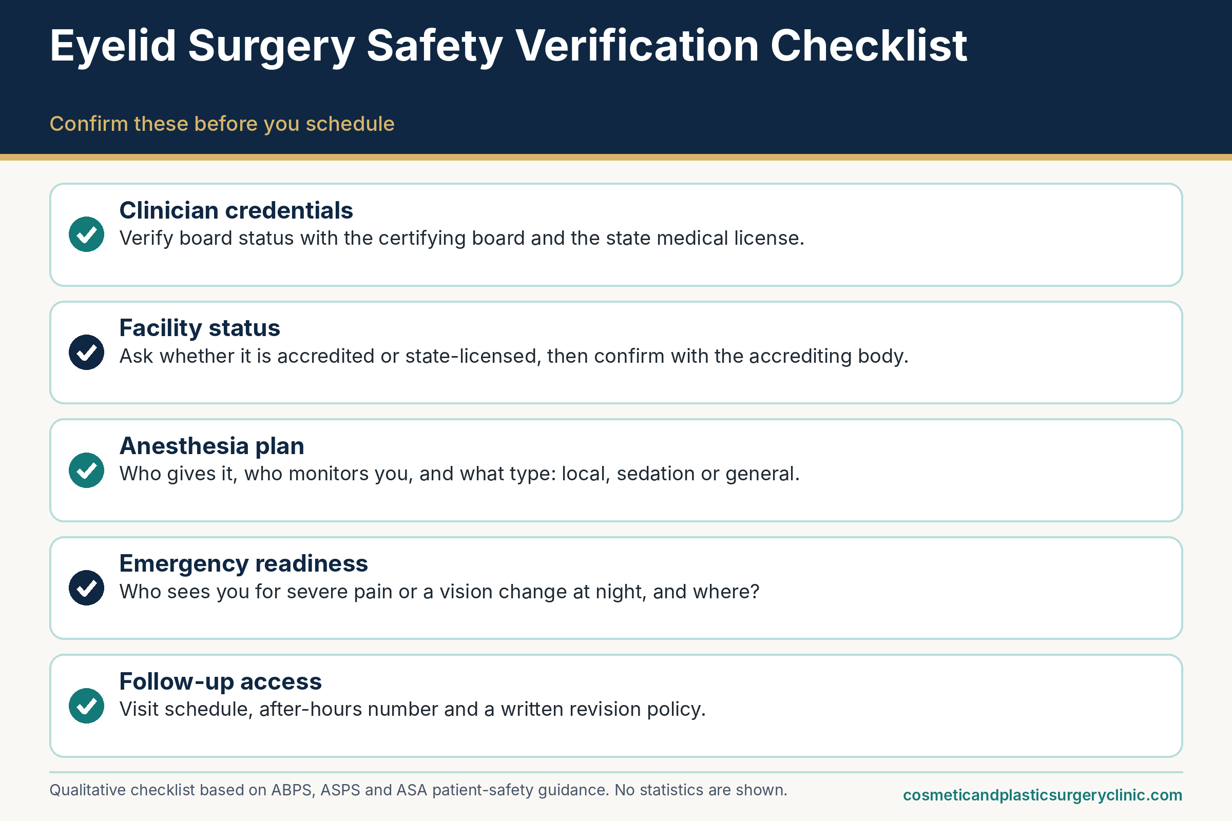
A risk-reduction pathway from consultation to follow-up
Risk cannot be eliminated, but a surprising share of it can be managed by process. Reading the surgical literature on eyelid complications, one theme recurs: prevention starts well before the first incision. The Brazilian single-center review says plainly that complication prevention begins with careful preoperative assessment, and Lelli and Lisman conclude that many problems are avoidable with planning and technique. This section lays out the pathway a thorough practice typically follows, so that patients can recognize it and notice what is missing. It describes common practice drawn from the sources reviewed; your own surgeon’s protocol governs, and it may differ.
Before surgery
The weeks before surgery are the best time to reduce risk, because this is when information can still change the plan, the timing, or the decision itself.
The pre-operative evaluation: history, eye examination, measurements and photographs
A good consultation for eyelid surgery is longer than a quick look in the mirror. Mayo Clinic describes a pre-operative workup that includes a review of surgical history and medical conditions, a complete eye examination, peripheral vision testing, and photography of the eyelids. Those elements each serve a purpose.
The history screens for conditions that raise risk: dry eye, previous eye or eyelid surgery, glaucoma, thyroid disease, circulatory problems, diabetes, bleeding tendencies and allergies. The eye examination checks that the eye itself is healthy and establishes a baseline. Lid measurements document how high the upper lid sits, whether the lids close completely, how loose the lower lids are, where the brow rests, and how symmetrical the starting point is. Tear production and tear film stability may be tested if dry eye is suspected. Peripheral vision testing documents whether drooping skin is limiting the visual field, which is relevant both for deciding whether surgery is functional and for later comparison. Photographs fix the starting point, a record that becomes useful later if there is a question about asymmetry, brow position or what changed.
The consultation is also where the surgeon decides whether eyelid surgery is the right operation at all. As noted earlier, ASPS observes that apparent lid drooping may reflect brow descent rather than lid excess, and that ptosis calls for a different procedure. A thoughtful evaluation therefore asks whether the problem is skin, fat, lid position, brow position, or something else entirely, and whether a different or additional procedure would fit better. Patients should expect to hear risks that are specific to their anatomy, not just a generic list, and to be asked about the expectations they bring. The guide to eyelid surgery candidacy goes into the logic of who is and is not evaluated for which approach.
Patients can prepare for this visit. Bring a list of every medication and supplement with doses, records of any previous eye or eyelid procedures, the name of your eye doctor if you have one, and a clear statement of what bothers you. If you have thyroid disease, dry eye disease, or an eye condition, bring documentation or ask your eye doctor to send a note. If you are considering surgery for vision reasons, ask what testing and photographs would be needed to document it.
Medication review, nicotine, blood pressure and practical arrangements
Once surgery is planned, the next phase is optimizing the factors that can be changed. The medication review should be explicit. Written instructions should say which medicines to continue, which to hold and for how long, and who made each decision. Mayo Clinic lists warfarin, aspirin, ibuprofen, naproxen and bleeding-promoting herbal supplements among the items to stop before surgery, with timing set by the surgeon, which is the general pattern. If you take any prescribed blood thinner, the plan should involve the prescriber, because stopping it without guidance can be hazardous.
Nicotine is the other change worth making early. Mayo Clinic advises quitting smoking several weeks before surgery, and the ASPS materials discussed earlier recommend avoiding all nicotine products, including e-cigarettes, for weeks before and after. If you smoke or vape and the timeline is short, tell the surgeon honestly. Practices handle this differently, but candor is the foundation of any plan, and a postponement is a small price compared with impaired healing.
Blood pressure is another item. If your blood pressure runs high, bring readings and ask whether it should be controlled before surgery and how it will be monitored during and after. A plan to prevent nausea and vomiting is a related safeguard, given the link between postoperative straining and bleeding discussed in the earlier section on bleeding behind the eye.
Finally, there are logistics that look mundane and still protect safety. Mayo Clinic advises arranging a ride home and having someone with you for the first night. Many practices also ask patients to stock lubricating ointment, cold compresses and dark sunglasses ahead of time, in line with the ASPS recovery guidance on lubrication, cold compresses and sun protection. Think about sleeping arrangements that allow the head to be elevated, a plan for pets and children, and time away from screens and strenuous work. Illustrative example: a person with a desk job who can work from home with short breaks has a lighter planning burden than someone whose work involves bending, lifting or dusty environments, who may need to arrange a longer leave. The guide to sleeping after eyelid surgery covers positioning in practical detail.
Putting these steps in sequence produces a planning calendar. The timing below is illustrative, assembled from the general patterns in the sources, and the instructions your surgeon gives always take priority. Several weeks ahead is the time for the consultation, any second opinion, and gathering records from earlier eye or eyelid procedures; it is also the time to start the nicotine break if the surgeon asks for one, since sources describe periods of several weeks. In the weeks before the date, the surgeon or an assistant reviews the medication and supplement list and tells you what to pause and when, and anyone with dry eye disease, thyroid disease or glaucoma has the relevant specialists involved. In the final week, the focus shifts to logistics: a ride home, a helper for the first night, ointment, cold compresses and sunglasses on hand, and a household arrangement that lets you rest with your head raised. The day before, you receive fasting and arrival instructions and confirm the after-hours number. After surgery, the calendar continues with a first check, commonly the next day or soon after, a suture-removal visit around the end of the first week if non-dissolving stitches were used, and later visits to check lid position and healing. A person who can see this whole calendar in advance is far less likely to find a missing piece at midnight.
Around surgery and afterward
Once the day arrives, risk reduction depends on teamwork, technique and the quality of the follow-up that follows.
Day of surgery: team, monitoring, positioning and technique choices
On the day, a few things should be visible. The team should confirm your identity, the planned operation and your medication and allergy information. The surgeon often marks the incision lines while you sit upright, since lid position changes when lying down. Monitoring of heart rhythm, blood pressure and oxygen levels should be continuous if you are sedated, and the person monitoring the sedation should have that as their focus, in line with the ASA statement discussed earlier. The eyes need protection during surgery, which can involve lubrication and protective shields; the 2012 review emphasizes lubrication during shield insertion and removal to avoid corneal abrasion.
Technique choices embody much of the risk mitigation described in the literature. These include conservative skin and fat removal, protecting the levator, meticulous control of bleeding, supporting the lower lid when laxity is present, and avoiding excessive lateral extension of incisions. The 2012 review summarizes key prevention strategies as thorough pre-operative assessment, precise measurements and symmetric planning, conservative fat and skin removal, levator protection, graded horizontal lid tightening in most lower-lid patients, appropriate suture selection and patient education on positioning and activity. Patients are not expected to supervise these steps, but they can ask the surgeon in plain language how they approach them.
Before discharge, the team should give written instructions with a phone number that is answered after hours, and should review the warning signs that mean “call now.” If the instructions list only a clinic number that goes to voicemail after five, ask what the after-hours plan is before the day of surgery, not after.
Aftercare, activity limits, follow-up visits and access to the team
Aftercare is risk management in a different form. The usual elements, consistent with the ASPS recovery page, Mayo Clinic and the 2012 review, include keeping the head elevated, applying cold compresses in the early period, using lubricating ointment or drops as directed, protecting the eyes from sun and wind, and avoiding rubbing, straining and heavy exertion until the surgeon allows it. The MedlinePlus entry gives one example of timelines, describing stitch removal within about five to seven days and a period of several weeks without strenuous activity, but individual instructions vary and yours take priority.
Follow-up visits serve a function beyond reassurance. At the first visit, usually within a day or so in many practices, the team can check visual acuity and the surface of the eye, as the 2012 review describes. At later visits, they check lid position, closure, wound healing and symmetry, and they remove sutures if needed. These checkpoints are where early malposition, exposure problems and scar changes are caught while they are still easy to manage, and they are the reason a practice’s follow-up policy deserves attention when comparing surgeons. The eyelid surgery recovery guide maps the typical sequence in more detail.
Patients contribute by following instructions, attending visits, taking dated photos if asked, and calling early rather than late. A common pattern in complication stories is a delay: a person who noticed something odd, wondered whether it was normal, and waited a day. The most important element of aftercare is a low threshold for calling. A good surgical team would rather take ten unnecessary calls than miss one real problem, and it is entirely appropriate to say so to the office at your pre-operative visit and ask them to confirm that calling is welcome.
Red flags: when to call the surgical team the same day and when to seek emergency care
The most useful safety tool after eyelid surgery is not a medication or a gadget. It is a clear idea of which symptoms can wait for the next business day, which deserve a call today, and which mean going to an emergency department right away. Patients often hesitate because they do not want to bother anyone or because they cannot tell whether what they feel is normal. This section offers a framework for thinking about escalation. It is general education, not triage, and it cannot replace the written instructions from your own surgeon. If you are unsure, treat the symptom as more urgent, not less.
The framework has three tiers. Emergency symptoms, which suggest a threat to vision or life, call for immediate emergency care or a call to 911. Same-day symptoms, which suggest a complication that needs evaluation but is not clearly an emergency, call for a phone call to the surgical team that day and, if you cannot reach anyone, escalation to urgent or emergency care. Routine questions, such as when you can resume exercise or why a small bump has appeared along the incision, can usually wait for office hours or the next scheduled visit.
Emergency-level symptoms
Two categories of symptoms call for immediate action: those suggesting pressure on the eye or optic nerve, and those suggesting a problem elsewhere in the body, such as a clot in the lung.
Sight-threatening symptoms: act immediately
As discussed in the section on bleeding behind the eye, the combination that should prompt urgent action is a change in vision, severe or escalating pain, and a tense, swollen, bulging eye. Mayo Clinic lists severe eye pain, bleeding and vision problems among the reasons to seek medical attention after blepharoplasty, and the clinical sources describe pain, proptosis, double vision, difficulty moving the eye and falling vision as features of orbital hemorrhage. The MedlinePlus entry on ectropion advises emergency attention for worsening vision, pain, light sensitivity or rapidly increasing redness.
In practical terms, many surgical teams give instructions along these lines, and yours may be similar or stricter: any sudden loss or dimming of vision, a dark curtain or missing area in the visual field, severe pain that is escalating or not relieved by the prescribed medication, a feeling of a tight, hard or bulging eye, inability to move the eye, or sudden one-sided swelling with pain should be treated as an emergency. Call 911 or go to the nearest emergency department, and notify the surgeon on the way if possible. Do not wait for a return phone call, and do not drive yourself if your vision is impaired.
When you arrive, state clearly that you recently had eyelid surgery and that you are worried about bleeding behind the eye or pressure on the optic nerve. That sentence helps the triage team prioritize and call an ophthalmologist or the on-call surgical service. If you can, bring your discharge instructions and the surgeon’s contact information. The reason for this urgency is time: as the orbital compartment syndrome review notes, visual outcomes are better the sooner pressure is relieved, and the window is measured in hours.
Whole-body emergencies: chest, breathing, clots and allergic reactions
Eyelid surgery involves anesthesia, medications and a period of reduced activity, and the usual emergencies of any surgery apply. Mayo Clinic includes shortness of breath, chest pain and abnormal heart rate in its list of reasons to seek medical attention after blepharoplasty. The CDC identifies difficulty breathing, chest discomfort that worsens with breathing, rapid or irregular heartbeat and fainting as warning signs of pulmonary embolism, and states that anyone with these symptoms should seek emergency care without delay. Those symptoms deserve a 911 call, not a message to the surgeon’s office.
Signs of a possible clot in a leg vein, such as new swelling, tenderness, warmth or discoloration in one leg or arm, call for prompt medical evaluation. The CDC describes DVT symptoms as warranting urgent, though not necessarily emergency, attention. Because many clots cause no symptoms at all, the CDC page is a reminder that absence of symptoms is not proof of absence of risk, but it is not a reason for alarm either.
Other emergencies include signs of a severe allergic reaction to a medication, such as widespread hives with swelling of the lips or tongue, wheezing, or trouble breathing, and new neurological symptoms like facial drooping, slurred speech, sudden weakness or confusion. None of these is specific to eyelid surgery, but they can occur in the recovery period, and the emergency response is the same as at any other time.
Same-day calls and routine questions
Most concerning symptoms after eyelid surgery fall into a middle zone. They are not clear emergencies, but they should not wait until the next appointment.
Concerns that warrant a same-day call
The following list reflects patterns described in the surgical and patient-education literature, and it is not exhaustive. Contact the surgical team the same day, including after hours, for:
- Swelling or bruising that is increasing, particularly if it is much greater on one side, after the early days when it should be leveling off.
- Pain that is not controlled by the prescribed regimen or that is getting worse rather than better.
- Bleeding that soaks through dressings or continues beyond mild oozing.
- Spreading redness, warmth, thick or foul drainage, a fever, or an incision that is opening.
- An eye that will not close comfortably, or a burning, gritty or sharp sensation that is increasing, especially with light sensitivity or a feeling that something is in the eye.
- Blurred vision that persists between ointment applications, or new double vision that does not clear.
- A lower lid that seems to be turning outward or pulling down, with tearing or exposure symptoms.
- Persistent or severe nausea, vomiting or coughing, which raise pressure around the eye and should be addressed promptly.
- Leg swelling, tenderness or calf pain, which warrants urgent evaluation as described above.
- A rash, itching or swelling that could signal a reaction to a medication or ointment.
The common thread is change and function. Symptoms that are getting worse over time, that interfere with eye closure or vision, or that arrive with pain or fever need attention soon. A typical surgical team would rather hear from a patient with a harmless concern than learn about a delayed problem at the next visit, and an office that discourages calls is a red flag in its own right.
Table 6 sets out the three tiers side by side, with examples. The flow diagram that follows presents the same thinking in a decision format.
| Tier | Examples | Suggested action | Why this tier |
|---|---|---|---|
| Emergency | Sudden vision loss or dark area; severe or escalating eye pain; tense, bulging eye; cannot move the eye; chest pain; difficulty breathing; fainting | Call 911 or go to the nearest emergency department now; notify the surgeon on the way | Possible pressure on the optic nerve, pulmonary embolism or another life- or sight-threatening event where minutes matter |
| Same-day call | Increasing one-sided swelling; uncontrolled pain; fever or spreading redness; wound opening; eye will not close; gritty, burning or light-sensitive eye that is worsening; persistent double vision; leg swelling | Call the surgical team that day, including after hours; escalate to urgent or emergency care if you cannot reach anyone or symptoms worsen | Possible infection, exposure injury, hematoma, lid-position problem or clot that needs examination soon |
| Routine question | Small bumps along the incision; mild itching; patchy numbness; questions about makeup, exercise, lenses, travel or scar care | Raise at the next visit or call during office hours | Common during healing and rarely time-sensitive, though anything that bothers you is worth asking |
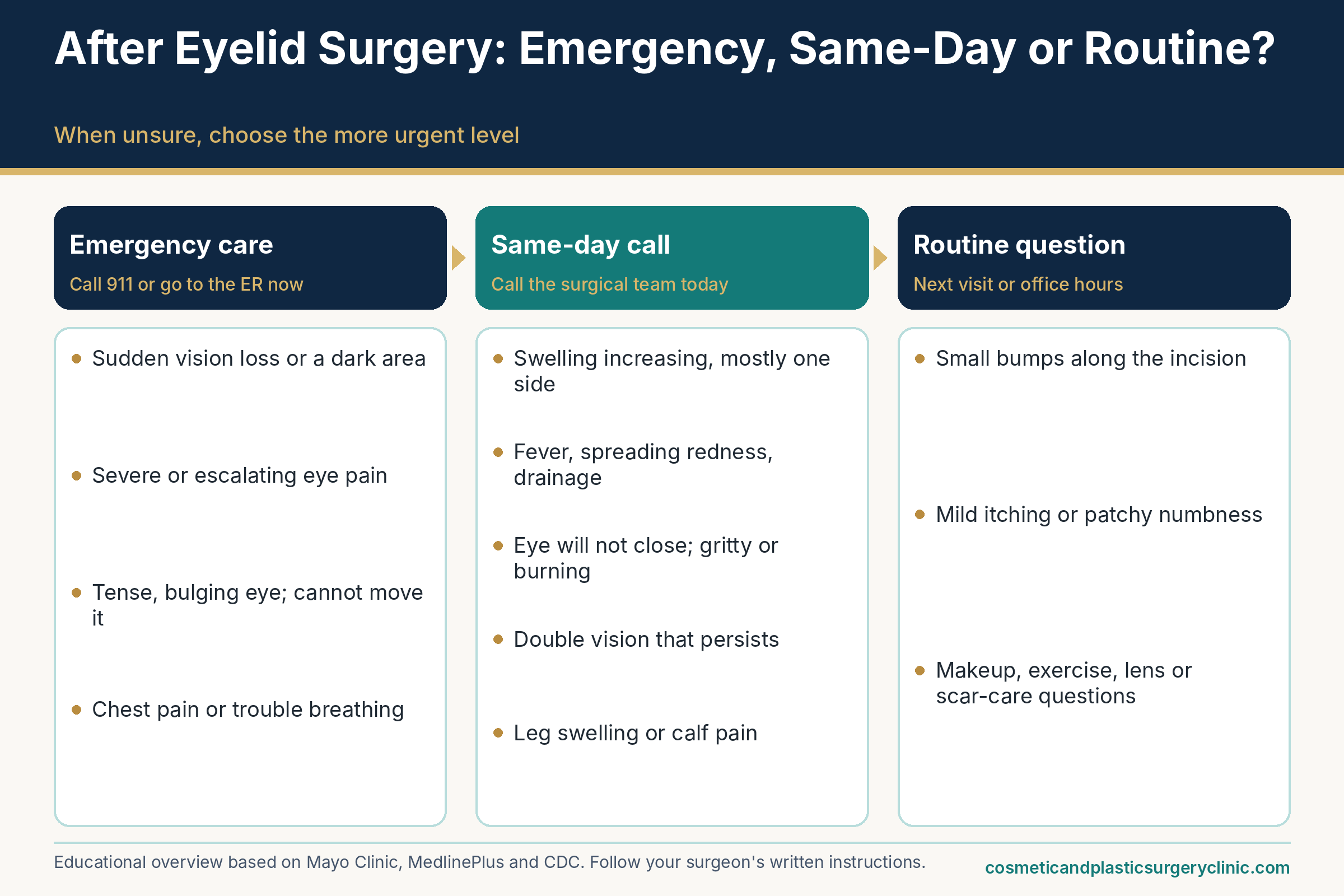
Four illustrative scenarios show how the framework works in practice. They are invented examples to illustrate reasoning, not real cases, and none of them substitutes for your team’s instructions. In the first, a person on the evening after surgery notices hazy vision for a few minutes after applying ointment, plus moderate and fairly equal swelling of both lids, with discomfort that is controlled by the prescribed regimen. That fits the expected pattern, and a note for the next check-in is enough. In the second, a person on day two finds that one upper lid is much more swollen and tight than the other, with an ache behind that eye that is growing, while vision seems normal for now. This is a same-day call without delay, and if the ache becomes severe or vision dims at any point while waiting for a callback, it becomes an emergency. In the third, a person on day eight sees redness spreading from one incision, warmth, yellowish drainage and a fever. That is also a same-day call, because infection needs examination, and the surgeon may want to see the person in the office that day. In the fourth, a person wakes at 3 a.m. with severe pain in one eye, a bulging sensation and a dark area in the visual field. That is an emergency: call 911 or go to the nearest emergency department immediately, and let the surgeon know when it is possible to do so. The difference between the second and fourth scenarios is speed and the presence of a vision change, and both look different from the first. When a situation seems to sit between two tiers, move up.
Making the call useful, and what to do if you cannot reach anyone
A call is more effective when you give the team a clear, brief picture. Say who you are, the date of surgery and the operation you had, what you are experiencing, when it started, whether it is changing, and what you have already tried or taken. Describe function (can you close your eye, can you see clearly) rather than only appearance. If it is safe to do so, take a clear photo in good light and offer to send it. Have your pharmacy and medication list on hand. Write down the instructions you are given and repeat them back.
If the call does not get through, do not assume the problem is minor. Practices should have a covering clinician or answering service for after-hours calls; if you reach a service, state that you have had eyelid surgery and describe your symptom clearly. If you cannot reach anyone and the symptom is in the emergency or same-day tier, go to an emergency department or urgent care, and ask them to contact your surgeon. If you are traveling, having surgery far from home, or having surgery abroad, ask in advance where you would be seen. The CDC and ASPS both note that follow-up and complication care for surgery done abroad can be difficult and costly, and that is worth weighing early. The guide on questions to ask about surgery abroad covers this in more detail.
A last note on mindset. Calling about a concern is not an admission of failure and it is not an imposition. Surgeons expect questions, and most complications that go well are caught early because the patient spoke up. Equally, a call that ends with reassurance is a good outcome. The goal is not to avoid unnecessary calls but to avoid unnecessary delay.
Revision, cost framing and functional versus cosmetic surgery
Not every unwelcome result is an emergency or even a clear complication. Sometimes the eyelids heal exactly as planned and the person still feels the change is too little, too much, or simply not what they pictured. Other times a true complication leaves the lids in a position that needs a second operation. This section covers how to think about revision, how to frame the money question without repeating the pricing guide, and how the line between functional and cosmetic surgery affects the risk-benefit balance.
Undercorrection, overcorrection and revision
Revision surgery has a reputation for being a quick “touch-up,” and sometimes it is. Often it is more involved than that, and the timing matters as much as the technique.
Telling undercorrection, overcorrection and ordinary healing apart
Undercorrection means the operation did not remove or reposition enough tissue to achieve the planned change. In the upper lid it might mean residual hooding or a heavy-looking fold. In the lower lid it might mean persistent puffiness. Overcorrection means too much was changed: too much skin or fat taken, a lid too high or too low, a hollow look, or a crease that sits too high. The 2006 Orbit review includes both over- and under-correction among the possible complications, and the 2012 Plastic Surgery International review spends considerable space on the consequences of excessive tissue removal, which include retraction, scleral show and lagophthalmos.
Overcorrection is generally the more serious of the two, because removed tissue cannot simply be put back and because it can compromise eye protection. Undercorrection is more often a matter of appearance and is usually easier to address, though it still means another operation. Both are different from the swelling-related, temporary versions of the same findings. A lid that looks heavy at one week may simply be swollen. A lid that looks too high may be held up by edema. The distinction is exactly why surgeons and the literature emphasize patience.
The 2012 review recommends delaying surgery to address persistent ptosis or minor asymmetries until at least three months after the operation so that swelling can resolve and scars mature, and it cautions against what it describes as chasing the result with early reoperation. That is a general principle, not a rule for every case. Problems that threaten the eye, such as persistent incomplete closure with corneal damage, may need earlier intervention, while appearance-driven revisions benefit from time. A frank discussion at the first postoperative visits about which category your concern falls into helps set a sensible timeline.
How common is revision? The honest answer is that the sources reviewed here do not provide a reliable population-level rate. In the Brazilian single-center series, 19 of 200 patients had a complication, and 37 percent of those patients needed revision surgery, a small absolute number from one center using one technique. That is the sort of figure that should not be extrapolated to other practices or to different operations. ASPS lists the possible need for revision surgery among the risks, so it belongs in the consent conversation for every patient. For readers who want to see how results are usually described over time, the eyelid surgery results guide and the guide to how long eyelid surgery results last discuss timelines and aging.
What revision surgery involves, and why it can be harder than the first operation
Revision work typically deals with altered anatomy: scar tissue, tissue shortage and disrupted planes. The techniques described in the literature reflect this. For lower-lid retraction or ectropion, the Healio discussion lists lateral tarsal strip procedures to tighten the lid, release of scar bands, spacer grafts using material such as hard palate mucosa, and skin grafts for severe shortage of the outer layer. For upper-lid retraction due to a shortage of skin, the 2012 review describes full-thickness skin grafts and other methods, and it notes that multiple repairs may be needed to reach the best result. For persistent ptosis, a posterior approach can raise the lid without reopening the original incision. For persistent asymmetry or webbing at the inner corner, minor scar revision techniques exist.
These descriptions are meant to give readers a sense of what the word “revision” can cover, not to suggest that any of these operations is routine or appropriate for a given person. They tend to involve donor sites (the skin from behind the ear or the lining of the roof of the mouth), added healing time, and results that can be good but not always ideal. Their complexity is the reason surgeons stress getting the first operation right and the reason it is sensible to consult a surgeon with specific experience in revision eyelid surgery if a problem occurs, even if that is not the person who did the original operation.
Practical steps that help any future surgeon include keeping the operative report, photographs from before and after surgery, a list of medications used, and notes about when problems began. If the first surgery was done elsewhere, request those records early. Revision decisions also benefit from honest reflection on whether the concern is functional (eye comfort, closure, vision) or aesthetic, because the urgency, the acceptable risk and the likely insurance position differ. A second opinion before a revision, ideally from a surgeon who regularly handles complex eyelid problems, is entirely reasonable and is not disloyal to the first surgeon.
Money and indications
Two financial and clinical frames deserve brief treatment here: how to think about the cost of complications and revision, and how functional versus cosmetic indications shape the risk-benefit picture.
How to frame the cost of complications and revision
This guide does not list prices; the eyelid surgery cost guide covers fees, price factors, insurance and financing, and the lower eyelid surgery cost guide covers the lower-lid specifics. What belongs in a safety guide is the structure of the question: who pays when something goes wrong, and what is the process?
The ASPS eyelid cost page states that most health insurance does not cover cosmetic surgery or its complications. That sentence is easy to read past, but it means that a complication following purely cosmetic surgery may generate out-of-pocket costs for office visits, medications, tests, procedures and perhaps facility and anesthesia charges for a second operation, along with time off work. Coverage depends on the plan and on the circumstances, so the point is to ask, not to assume. Some complications that affect eye health may be handled by health insurance as medical care, but that is a plan-specific matter outside what a general article can say.
Before booking, ask for the practice’s revision and complication policy in writing. A useful policy answers specific questions. Does it apply to the surgeon’s fee only, or also to the facility and anesthesia charges? How long does it last? Does it cover complications, revisions for appearance, or both? Under what conditions is it void? What happens if you need care from another physician, for instance an ophthalmologist for a corneal problem? An assurance of “free touch-ups” in marketing language is not a policy, and it is not a safety measure. A low price with a vague complication plan can cost more than a higher price with clear terms.
Surgery abroad adds a distinct set of financial risks. The ASPS briefing paper on cosmetic surgery tourism warns that complications and revision costs can add up to more than the original surgery would have cost in the U.S., that following up on problems at home can be difficult, and that there may be no legal recourse after negligence. The CDC’s guidance on medical tourism likewise notes that treating complications back in the U.S. can be expensive and may not be covered by insurance. These points apply with particular force to any operation near the eye, where early access to an experienced surgeon matters.
Functional versus cosmetic indications, briefly
Eyelid surgery serves two broad purposes. In the cosmetic case, the goal is appearance: a more rested look, less hooding, smoother lids. In the functional case, the goal is to relieve a medical problem, usually upper-lid skin that droops far enough to block part of the visual field or rest on the lashes. The MedlinePlus entry notes that surgery is needed when drooping eyelids obstruct vision, and that some people pursue it for cosmetic improvement. The ASPS cost page says that when eyelid surgery removes excess skin that obstructs the eyelashes, it may be eligible for insurance reimbursement, and that policies should be reviewed carefully. Mayo Clinic notes that the pre-operative workup includes peripheral vision testing.
Does the distinction change risk? The surgical risks are broadly the same for the same operation, but the balance with benefit differs. When a real functional limitation exists, the benefit side of the ledger is larger and a patient may reasonably accept a risk that would be less justified for a purely aesthetic goal. When the motivation is cosmetic, a person can weigh the risks against a goal that is personal and discretionary, and can reasonably choose to wait, choose a less invasive alternative, or decide against surgery. Neither frame is better or worse; the point is that the risk conversation should be tailored to the reason for surgery.
Two cautions apply. First, insurance coverage is not a quality indicator, and a procedure being “medically necessary” does not mean it is simpler or safer. Second, payers have their own documentation requirements, which can include photographs and visual field testing, and the details differ from one plan to the next. The cost guide linked above covers that process. For lower-lid concerns, functional indications are less common, which is another reason risk-benefit analysis for lower-lid surgery tends to rest more on personal goals. Readers weighing nonsurgical or less invasive paths can compare them in the guide to alternatives to eyelid surgery.
Informed consent, choosing a surgeon and separating myth from fact
Everything in this guide converges on two moments: the consent conversation, when you decide whether to proceed knowing the material risks, and the choice of who will operate. Both are better done slowly. Neither is a formality. This final main section describes what a good consent process contains, how to verify the credentials of different kinds of eyelid surgeons without declaring any one path superior, and how to sort the claims you will encounter online into those that hold up and those that do not.
Informed consent
Informed consent is often reduced to a signature. Ethically and practically it is a conversation that the signature records.
What a good consent discussion covers
The American Medical Association’s Code of Medical Ethics describes informed consent as a process in which the physician explains the diagnosis when known, the nature and purpose of the recommended intervention, the burdens, risks and expected benefits of the options, and the alternatives, including no treatment, in a way that fits the patient’s preferences for receiving information. It also calls for assessing the patient’s ability to understand and make a voluntary decision, and for documenting the discussion in the record. ASPS adds, on its eyelid safety page, that patients must sign consent documents confirming that they understand the risks and complications, and it advises raising all questions directly with the plastic surgeon.
For eyelid surgery, a thorough consent discussion goes beyond a generic list and ought to address the following, in plain language and with room for questions.
The specific operation planned. Which lids, which technique, whether fat will be removed or repositioned, whether the lower lid will be tightened, and whether anything else is being done at the same time. The name on the consent form should match the plan you were quoted.
The material risks, tailored to you. This means the ones most likely to matter in your case, such as dry eye and exposure if you already have a dry eye history, lid malposition if your lower lids are lax, or bleeding if you take a blood thinner, alongside the rare serious risks. A surgeon should be willing to say plainly that bleeding behind the eye can threaten vision, even though it is rare, and what the plan is if it happens.
What is expected versus what is a complication. The surgeon should explain what the early recovery will look and feel like and what warrants a call, ideally with written instructions that include an after-hours number.
Alternatives, including doing nothing. Alternatives may include brow lift, ptosis repair, nonsurgical treatments for under-eye hollows, skin-tightening treatments or watchful waiting. Even if you prefer surgery, hearing the alternatives is part of consent.
Who will do what. Who performs the surgery, who administers anesthesia, who is in the room, where the surgery will take place, and who is available if something goes wrong.
Revision and complication policy. What happens if a second procedure is considered, how costs are handled, and who decides, ideally written down.
Use of products or techniques outside their labeled use. If a laser, filler, injectable or medication is part of the plan, ask whether its use here is on-label or off-label. The Food and Drug Administration explains that off-label use means using an approved product for an unapproved purpose, dose or route; clinicians generally may do this when they judge it medically appropriate, but the FDA has not determined safety and effectiveness for that use. Off-label use is not inherently improper and is common in medicine. The relevant consent point is that you should be told, and that the surgeon should explain why, what the evidence is, and what the alternatives are.
Expectations, photographs, alternatives and the right to pause
Complication rates tell only part of the satisfaction story. A person who has an uncomplicated operation but expected a different outcome may still feel let down. For this reason, expectation-setting is a safety measure as much as a courtesy. A conscientious surgeon will use your pre-operative photographs to point out existing asymmetries, discuss the difference between a subtle and a dramatic change, and explain what eyelid surgery does not address, such as wrinkles, brow position or dark circles, as MedlinePlus points out in its description of the operation’s limits. The ASPS candidates page makes the related point that eyelids are part of the overall facial anatomy and that a qualified plastic surgeon evaluates the whole face rather than assuming that all eyelid concerns need the same procedure.
Equally important is the right to pause. ASPS describes the decision to undergo eyelid surgery as extremely personal, to be weighed against the risks. Nothing about consent requires you to decide on the day of the consultation. A practice that hurries you toward a deposit, offers a discount that expires today, or discourages a second opinion is behaving in ways that sit uneasily with informed consent. Taking a few days or weeks to review your questions, compare a second consultation, and reread the consent form at home is ordinary and sensible. The guide to eyelid surgery consultation questions and the natural-looking results guide are good companions for this stage.
A second opinion deserves a few words, because people sometimes feel it is disloyal or a waste of time. It is neither. A second consultation lets you compare how two surgeons assess the same lids, whether they recommend the same operation and, just as important, whether they describe the same risks. If two surgeons agree about the plan but describe the risks very differently, that gap is worth exploring. If they disagree about the plan, the difference can clarify the trade-offs. Bring the same list of questions to both, take notes, and compare answers about emergencies and follow-up as carefully as answers about technique. Most reputable surgeons expect patients to seek more than one opinion.
Choosing a surgeon and separating myth from fact
The goal of this subsection is not to name a winner among specialties. It is to help you verify what you are told and ask questions that reveal experience.
Credentials and training paths, verified without ranking
Eyelid surgery in the United States is performed by clinicians from several training backgrounds. The sources reviewed here describe the verification tools, not comparative outcomes, and this review located no verified evidence that one pathway produces better eyelid outcomes than another. What matters is that each credential is real, current and relevant to the operation.
For plastic surgeons, the American Board of Plastic Surgery (ABPS) offers a public verification tool that searches by name or location. ABPS explains that certification reflects completion of appropriate training and passing written and oral examinations, that an active, unrestricted medical license is required, and that if a state medical board has taken action against a certified surgeon an alert directs users to the Federation of State Medical Boards. ASPS adds that board certification is voluntary, outlines the training path to ABPS certification, and warns that some “board certifications” come from boards not recognized by the American Board of Medical Specialties or are in unrelated specialties. The American Board of Medical Specialties maintains a free “Certification Matters” lookup that covers its member boards, which lets you check a surgeon’s certification in any specialty they claim. The guide to ABPS board certification and the verification checklist walk through the steps.
For oculoplastic (oculofacial plastic) surgeons, the professional society, the American Society of Ophthalmic Plastic and Reconstructive Surgery (ASOPRS), keeps a member directory and a fellowship directory, and describes fellowship training in surgery of the eyelids and surrounding structures. Society membership is not the same as board certification, so the ophthalmologist’s primary board status should be verified separately. For facial plastic surgeons, the American Board of Facial Plastic and Reconstructive Surgery, established in 1986, offers a surgeon finder to confirm certification; its website does not state whether it is a member of the American Board of Medical Specialties, so check that point with ABMS rather than assume it. In all cases, a state medical license is a separate requirement that can be verified through the state medical board.
| What you are checking | Where to check | What it confirms | What it does not confirm |
|---|---|---|---|
| ABPS board certification in plastic surgery | ABPS online verification tool | Current certification status; active unrestricted license is a prerequisite | Experience with a particular eyelid technique or complication rates |
| Board certification in any ABMS specialty (for example, an ophthalmologist or other physician) | ABMS Certification Matters lookup | Certification by an ABMS member board in the named specialty | Eyelid-specific training or fellowship |
| Oculoplastic or oculofacial fellowship training | ASOPRS member and fellowship directories; ask the surgeon directly | Society membership or listed fellowship training | Primary board certification; individual outcomes |
| Facial plastic surgery certification | American Board of Facial Plastic and Reconstructive Surgery surgeon finder; confirm board recognition status with ABMS if relevant | Certification by that board | Whether the board is ABMS-recognized, unless separately verified |
| State medical license and disciplinary history | State medical board website; Federation of State Medical Boards alerts | Legal authority to practice; any public actions | Specialty training or board status |
| Facility accreditation or licensure | AAAHC, AAAASF, Joint Commission or state listing; Medicare certification | Facility meets that body’s standards | The quality of any individual surgeon or anesthesia provider |
Credentials answer “Is this person qualified in general?” They do not fully answer “Is this person right for my eyelids?” The questions in the next subsection help with the second.
Questions to ask, myths versus facts, and red flags in marketing
Table 8 organizes consultation questions by what they reveal. You do not need to ask all of them; choose those that fit your situation, and listen for specific answers rather than reassurance. For a broader list, see the 25 questions to ask a plastic surgeon.
| Topic | Question to ask | What a useful answer sounds like | Cause for concern |
|---|---|---|---|
| Your anatomy | What did you find about my lid laxity, tear production and brow position, and how does that change the plan? | Specific findings and an explanation of how they shape the technique | A generic plan that ignores examination findings |
| Risks that apply to me | Which complications are most relevant for me, and which are rare but serious? | Tailored list that includes dry eye, lid position and bleeding behind the eye | Dismissing risks as “very rare” with no detail |
| Technique | Which technique for each lid, and why? Will the lower lid be tightened? | Reasons tied to your skin, fat and lid support | One approach offered to everyone |
| Experience | How often do you perform this operation, and how do you handle lid malposition or dry eye? | Clear description of experience and a management approach | Defensiveness or vague answers |
| Anesthesia and facility | Who will give anesthesia and monitor me? Where is the surgery, and how is it accredited or licensed? | Named role and credentials; accreditation or license you can verify | Unable or unwilling to say who monitors sedation |
| Emergencies | If I had severe pain or vision change overnight, who would I call and where would I be seen? | A direct number, a covering clinician and a hospital plan | “Just go to the emergency room” with no coordination |
| Follow-up | What follow-up visits are included, and what is your revision policy in writing? | A defined schedule and written policy | Verbal promises of free touch-ups |
| Alternatives and decision pace | What are my alternatives, and what happens if I wait? | Honest options, including nonsurgical choices and waiting | Pressure to decide or pay a deposit today |
The questions in Table 8 can also be summarized on one page to take with you.
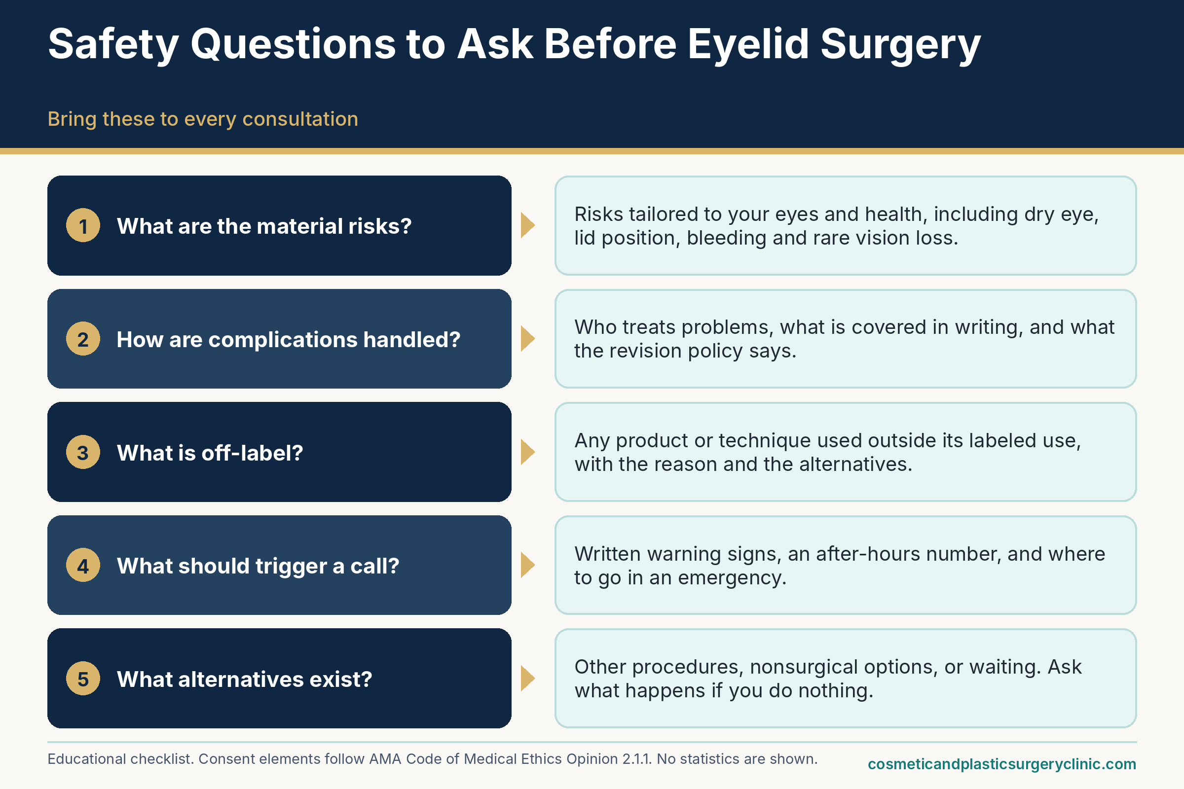
Marketing deserves its own skepticism. Language to treat with caution includes claims that surgery carries no risk or is “painless,” promises of results that are “scarless” or “permanent” without qualification, discounts that expire within days, and galleries of before-and-after photos with no information about lighting, timing, technique or whether the pictured patients had similar anatomy. The guide to evaluating eyelid before-and-after photos explains how to read them. Reviews and social media posts are not evidence of safety: they cannot tell you about complication rates, and a single glowing or disastrous story does not describe your odds.
Table 9 tests some common beliefs against the sources reviewed here.
| Common belief | What the sources support | Why it matters |
|---|---|---|
| “It is a minor procedure, so there is nothing serious to worry about.” | Most recoveries are uneventful, but ASPS and Mayo Clinic list real risks, including rare vision loss. | Common and short does not mean trivial; consent should cover the rare serious risks |
| “Any licensed doctor can do it equally well.” | A license is the legal minimum; board certification and specific eyelid experience are separate considerations that can be verified. | Planning and technique drive many preventable complications |
| “Swelling and bruising are all that happens.” | Those are the usual effects, but complications also include dry eye, lid malposition, asymmetry, scarring and infection. | Knowing the full list helps you recognize what is not normal |
| “A lower price means the same operation for less.” | Quotes can differ in surgeon, anesthesia, facility, scope and follow-up; ASPS notes cosmetic complications are generally not covered by insurance. | Compare scope and safeguards, not totals alone |
| “If something goes wrong, revision will simply fix it.” | Revision is often possible but can be more complex than the first operation and may involve grafts and further healing. | The first operation is the best time to get the plan right |
| “Dry eyes after surgery always go away quickly.” | Often temporary, but a 2021 study found pre-existing dry eye linked to more persistent surface changes. | Existing dry eye should be evaluated before surgery |
| “Smoking only matters for bigger operations like facelifts.” | Mayo Clinic advises quitting smoking weeks before blepharoplasty, and ASPS lists non-smoking among candidate characteristics; nicotine in any form is a concern. | Nicotine affects blood flow and healing in any operation; follow your surgeon’s timeline |
| “Vision loss is something I could not do anything about.” | Bleeding behind the eye is time-sensitive; earlier recognition and treatment are linked to better outcomes in the literature. | Knowing the warning signs and having an emergency plan is protective |
One way to hold all of this: the best-informed decisions tend to share three features. The person understands what they are accepting. The team is verifiably qualified and the setting is suitably equipped. And there is a clear plan for the unlikely event, including who to call. None of those features requires an ideal surgeon or an ideal patient. They require a conversation that is complete enough to surface what matters, and the time to have it.
Frequently asked questions about blepharoplasty risks and complications
Is eyelid surgery safe?
Safe is a relative word here. Most people who have eyelid surgery recover without a serious problem, but the operation carries real risks, including dry eye, lid-position changes, scarring, bleeding, infection, anesthesia reactions and, very rarely, vision loss. How risky it is for you depends on which lids are treated, your eye and health history, the anesthesia plan, the facility and the surgeon. A careful consultation can describe your risks more specifically than any general article. Treat any source that promises no risk with caution.
What is the most common complication after blepharoplasty?
No source reviewed for this guide ranks complications by frequency across all surgeons, so a definitive answer is not available. The 2012 review in Plastic Surgery International describes lid-height and crease asymmetry as the outcome patients most commonly notice, and bruising as universal. In the single-center Brazilian series, chemosis (swelling of the membrane over the white of the eye) and lower-lid malposition were among the more frequent problems. Frequency depends heavily on the technique, the patient group and how strictly minor problems are counted, so ask a surgeon for figures from their own practice.
How likely is vision loss from eyelid surgery?
Very unlikely, but not impossible. A 2004 survey of ophthalmic plastic surgeons covering 269,433 cosmetic eyelid cases reported orbital hemorrhage in about 1 in 2,000 and permanent visual loss in about 1 in 22,000, according to its abstract. Those figures rely on surgeon recall and are two decades old, so they are best read as a rough order of magnitude. Because the consequences are serious, know the warning signs, severe pain, bulging and falling vision, and ask the surgeon what the emergency plan is.
How long do dry or watery eyes last after eyelid surgery?
It varies. In a small 2021 study of 55 young women after upper-lid surgery, blink changes returned to baseline within a month. Another small 2021 study found that people with healthy eyes had tear-stability changes that recovered by six months, while those with existing dry eye showed more persistent changes. Existing dryness may lengthen the course, and the sources reviewed do not quantify how age or lower-lid surgery affect it. Report dryness that is worsening, painful or accompanied by blurred vision, because the surface of the eye can be injured.
Is lower eyelid surgery riskier than upper eyelid surgery?
The risks differ more than they rank. Upper-lid surgery concentrates risk around eye closure, lid height and crease asymmetry. Lower-lid surgery concentrates it around lid support, with retraction, scleral show and ectropion discussed most. The 2006 Orbit review describes lower-lid problems as among the most significant aesthetic concerns, but no verified comparison of overall risk was found for this guide, and the right operation depends on anatomy and goals. A surgeon who explains which structures are at issue in your case is giving you more useful information than a general ranking.
Could I be left unable to close my eyes?
It is a recognized complication, called lagophthalmos, and most often follows removal of too much skin, scarring or lid retraction. Temporary incomplete closure from swelling is more common and typically improves as swelling subsides. Closure that remains incomplete after swelling has resolved, or that causes burning, redness or light sensitivity, warrants prompt evaluation because the cornea can be damaged. Treatment ranges from lubricating drops and ointment to taping at night and, for persistent cases, surgery to lengthen or reposition the lid.
How can I tell normal bruising from a dangerous collection of blood?
Ordinary bruising is spread out, may drift down the cheek, and tends to stabilize and then fade. A concerning pattern is swelling that is tense, markedly one-sided and still increasing, especially with pain that is out of proportion, a bulging eye, or any change in vision. A small collection confined to the eyelid skin is different from bleeding behind the eye, and only an examination can tell them apart. If you are unsure, call the surgical team the same day; if vision changes or pain is severe, seek emergency care.
Can I have eyelid surgery if I have dry eye disease or thyroid eye disease?
Possibly, but both conditions change the planning. Surgeons commonly evaluate and treat dry eye first and may suggest a more conservative operation or postponement. In thyroid eye disease, eyelid surgery is usually considered after the disease has become stable and after any orbital or eye-alignment surgery, according to a review in Frontiers in Endocrinology. Either condition should be disclosed at the first visit, with records from your eye doctor or endocrinologist if you have them, so the surgeon can decide whether and when surgery makes sense.
Do I need to stop blood thinners or supplements before eyelid surgery?
Surgeons usually ask patients to pause certain medications and supplements before surgery, and Mayo Clinic names warfarin, aspirin, ibuprofen, naproxen and bleeding-promoting herbal products. But whether and when to stop depends on why you take them. Prescribed anticoagulants are often protecting against stroke or clots, so do not stop anything on your own. Bring a complete list to the pre-operative visit, and let the surgeon and the prescribing clinician decide together. If pausing is unsafe, postponing the elective operation may be the better choice.
When can a revision or touch-up be considered?
For minor asymmetry or mild ptosis, a 2012 review advises waiting at least three months so that swelling subsides and scars mature. Problems that endanger the eye, such as incomplete closure with corneal injury, may need earlier action. Before any revision, ask the original surgeon for a candid assessment, keep your operative records and photographs, and consider a second opinion from a surgeon experienced in revision eyelid work. Also ask whether your revision policy covers facility and anesthesia charges, not just the surgeon’s fee.
Will insurance cover complications from cosmetic eyelid surgery?
Often not. ASPS states that most health insurance does not cover cosmetic surgery or its complications. Coverage depends on your plan, on whether the original surgery was functional or cosmetic, and on how the plan classifies the complication. Do not assume; ask the practice what costs would be yours, and ask your insurer before surgery if you can. Functional upper-lid surgery documented for vision problems may be handled differently, which the cost guide on this site explains.
Does general anesthesia make eyelid surgery riskier than local anesthesia with sedation?
Each option brings different trade-offs, and none is automatically safer. General anesthesia secures the airway but adds drug and airway risks and more nausea. Local anesthesia with sedation avoids deep anesthesia, though sedation can become deeper than intended and local injection near the eye has its own small risks. What matters most is who gives and monitors it, the facility’s readiness, and your health. The American Society of Anesthesiologists distinguishes monitored anesthesia care from moderate sedation and emphasizes qualified anesthesia providers, especially in office-based settings.
Does vaping or nicotine replacement affect eyelid surgery?
Nicotine in any form can constrict blood vessels, and ASPS materials from 2016 advise avoiding all nicotine products, including e-cigarettes, gum and patches, for weeks before and after plastic surgery. The ASPS e-cigarette release described the direct evidence on vaping and surgical outcomes as limited while calling a four-week break prudent. Because the exact timeline is set by your surgeon, disclose all nicotine use at the first visit. Nicotine also worsens Graves’ eye disease, which matters if you have a thyroid history.
Should I see an eye doctor before eyelid surgery?
Mayo Clinic describes a complete eye examination, peripheral vision testing and eyelid photography as part of the preparation. An additional visit with your own ophthalmologist or optometrist is especially reasonable if you have dry eye, glaucoma, thyroid eye disease, previous eye surgery such as LASIK, or limited vision in one eye. They can document your baseline and coordinate with the surgeon about drops, testing and timing. Ask your surgeon whether they want a note or records beforehand.
Sources and further reading
- American Society of Plastic Surgeons — Eyelid surgery: safety and risks (accessed 2026-10-03) — listed risks and consent
- American Society of Plastic Surgeons — Eyelid surgery: candidates (accessed 2026-10-03) — candidate characteristics, brow descent and ptosis
- American Society of Plastic Surgeons — Eyelid surgery: procedure (accessed 2026-10-03) — anesthesia, upper and lower approaches, sutures
- American Society of Plastic Surgeons — Eyelid surgery: recovery (accessed 2026-10-03) — expected early effects, sun protection
- American Society of Plastic Surgeons — Eyelid surgery: cost (accessed 2026-10-03) — insurance and complications
- Mayo Clinic — Blepharoplasty (accessed 2026-10-03) — risks, preparation, when to seek care
- MedlinePlus — Eyelid lift surgery (page updated February 5, 2025; accessed 2026-10-03) — risks, recovery, limits of the operation
- MedlinePlus — Ectropion (information current July 9, 2024; accessed 2026-10-03) — outward-turning lid, symptoms, urgent care
- Patrocinio et al. — Complications in blepharoplasty: how to avoid and manage them, Brazilian Journal of Otorhinolaryngology (accessed 2026-10-03) — single-center retrospective series of 200 patients; prevention and management
- Oestreicher and Mehta — Complications of blepharoplasty: prevention and management, Plastic Surgery International, 2012 (accessed 2026-10-03) — review of complications and revision timing
- Incidence of postblepharoplasty orbital hemorrhage and associated visual loss, Ophthalmic Plastic and Reconstructive Surgery, 2004 (abstract; accessed 2026-10-03) — survey figures for hemorrhage and visual loss
- Lelli and Lisman — Blepharoplasty complications, Plastic and Reconstructive Surgery, 2010 (abstract record; accessed 2026-10-03) — complications organized by timing
- Complications of blepharoplasty, Orbit, 2006 (PubMed abstract; accessed 2026-10-03) — overview of complication types
- McCallum et al. — Orbital compartment syndrome: an update with review of the literature, Clinical Ophthalmology, 2019 (accessed 2026-10-03) — causes, signs, time sensitivity, canthotomy
- American Academy of Ophthalmology EyeWiki — Retrobulbar hemorrhage (accessed 2026-10-03) — symptoms, timing, risk factors, management
- StatPearls — Lagophthalmos (Fu and Patel, 2023; accessed 2026-10-03) — incomplete closure and corneal exposure
- Healio — How do I manage lower eyelid malposition following lower eyelid blepharoplasty? (September 30, 2020; accessed 2026-10-03) — causes and management of lower-lid malposition
- Frontiers in Medicine — Effect of transcutaneous upper eyelid blepharoplasty on blink parameters and lipid layer thickness (2021; accessed 2026-10-03) — small prospective study of blink changes
- Frontiers in Medicine — Changes of dry eye related markers and tear inflammatory cytokines after upper blepharoplasty (2021; accessed 2026-10-03) — pre-existing dry eye and persistence of surface changes
- OphthalmologyWeb — Guidelines for performing blepharoplasty in post-LASIK patients (summary of Korn et al., 2007; accessed 2026-10-03) — waiting period and dry eye testing
- National Eye Institute — Dry eye (accessed 2026-10-03) — symptoms, risk factors, treatment
- National Eye Institute — Graves eye disease (accessed 2026-10-03) — symptoms and surgical options
- NIDDK — Graves’ disease (last reviewed November 2021; accessed 2026-10-03) — prevalence, eye disease, smoking
- Frontiers in Endocrinology — Update on the surgical management of Graves’ orbitopathy (accessed 2026-10-03) — order of surgeries and timing
- Centers for Disease Control and Prevention — Venous thromboembolism (accessed 2026-10-03) — clot risk factors and warning signs
- American Society of Plastic Surgeons — How nicotine sabotages plastic surgery (December 12, 2016; accessed 2026-10-03) — nicotine and healing
- American Society of Plastic Surgeons — E-cigarettes and plastic surgery (November 28, 2016; accessed 2026-10-03) — e-cigarette guidance
- American Society of Anesthesiologists — Statement distinguishing monitored anesthesia care from moderate sedation/analgesia (October 2023; accessed 2026-10-03) — anesthesia care definitions
- American Society of Anesthesiologists — Statement on qualifications of anesthesia providers in the office-based setting (reaffirmed October 2024; accessed 2026-10-03) — office-based anesthesia
- American Society of Plastic Surgeons — Accredited facilities (accessed 2026-10-03) — accrediting bodies and standards
- American Society of Plastic Surgeons — Why board certification matters for plastic surgery (March 26, 2025; accessed 2026-10-03) — training path and credential cautions
- American Board of Plastic Surgery — Verify certification (accessed 2026-10-03) — public verification tool
- American Board of Medical Specialties — Certification Matters (accessed 2026-10-03) — lookup across member boards
- American Society of Ophthalmic Plastic and Reconstructive Surgery (accessed 2026-10-03) — member and fellowship directories
- American Board of Facial Plastic and Reconstructive Surgery (accessed 2026-10-03) — surgeon finder
- American Medical Association — Code of Medical Ethics Opinion 2.1.1, Informed consent (accessed 2026-10-03) — elements of informed consent
- U.S. Food and Drug Administration — Understanding unapproved use of approved drugs “off label” (page last modified February 5, 2018; accessed 2026-10-03) — meaning of off-label use
- Centers for Disease Control and Prevention — Medical tourism (accessed 2026-10-03) — risks of surgery abroad and follow-up
- American Society of Plastic Surgeons — Briefing paper: cosmetic surgery tourism (accessed 2026-10-03) — credentials, follow-up and complication costs